Deposition Date
2011-09-02
Release Date
2011-11-23
Last Version Date
2023-12-20
Entry Detail
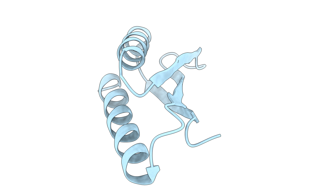
PDB ID:
3ZZP
Keywords:
Title:
Circular permutant of ribosomal protein S6, lacking edge strand beta- 2 of wild-type S6.
Biological Source:
Source Organism(s):
THERMUS THERMOPHILUS (Taxon ID: 274)
Expression System(s):
Method Details:
Experimental Method:
Resolution:
0.96 Å
R-Value Free:
0.13
R-Value Observed:
0.11
Space Group:
P 21 21 21