3ZYV image
Deposition Date 2011-08-27
Release Date 2012-09-05
Last Version Date 2023-12-20
Entry Detail
PDB ID:
3ZYV
Keywords:
Title:
Crystal structure of the mouse liver Aldehyde Oxidase 3 (mAOX3)
Biological Source:
Source Organism:
MUS MUSCULUS (Taxon ID: 10090)
Method Details:
Experimental Method:
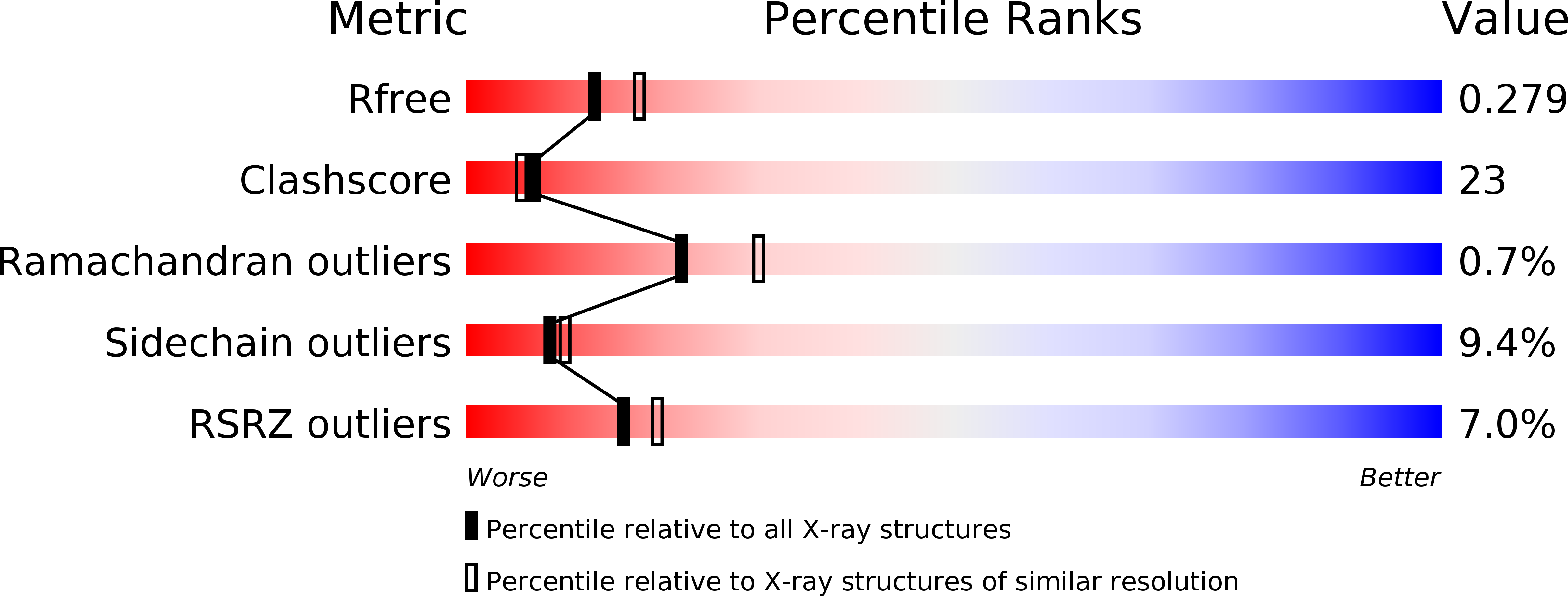
Resolution:
2.55 Å
R-Value Free:
0.28
R-Value Work:
0.25
R-Value Observed:
0.25
Space Group:
P 1
Macromolecular Entities
Polymer Type:polypeptide(L)
Molecule:AOX3
Chain IDs:A, B, C, D
Chain Length:1335
Number of Molecules:4
Biological Source:MUS MUSCULUS
Primary Citation
The First Mammalian Aldehyde Oxidase Crystal Structure: Insights Into Substrate Specificity.
J.Biol.Chem. 287 40690 ? (2012)
PMID: 23019336 DOI: 10.1074/JBC.M112.390419

Abstact

BACKGROUND Aldehyde oxidases have pharmacological relevance, and AOX3 is the major drug-metabolizing enzyme in rodents. RESULTS The crystal structure of mouse AOX3 with kinetics and molecular docking studies provides insights into its enzymatic characteristics. CONCLUSION Differences in substrate and inhibitor specificities can be rationalized by comparing the AOX3 and xanthine oxidase structures. SIGNIFICANCE The first aldehyde oxidase structure represents a major advance for drug design and mechanistic studies. Aldehyde oxidases (AOXs) are homodimeric proteins belonging to the xanthine oxidase family of molybdenum-containing enzymes. Each 150-kDa monomer contains a FAD redox cofactor, two spectroscopically distinct [2Fe-2S] clusters, and a molybdenum cofactor located within the protein active site. AOXs are characterized by broad range substrate specificity, oxidizing different aldehydes and aromatic N-heterocycles. Despite increasing recognition of its role in the metabolism of drugs and xenobiotics, the physiological function of the protein is still largely unknown. We have crystallized and solved the crystal structure of mouse liver aldehyde oxidase 3 to 2.9 Å. This is the first mammalian AOX whose structure has been solved. The structure provides important insights into the protein active center and further evidence on the catalytic differences characterizing AOX and xanthine oxidoreductase. The mouse liver aldehyde oxidase 3 three-dimensional structure combined with kinetic, mutagenesis data, molecular docking, and molecular dynamics studies make a decisive contribution to understand the molecular basis of its rather broad substrate specificity.

Legend

Protein

Chemical

Disease

Primary Citation of related structures