Deposition Date
2011-06-28
Release Date
2011-12-14
Last Version Date
2023-12-20
Entry Detail
PDB ID:
3ZSL
Keywords:
Title:
Crystal structure of Apo Human Galectin-3 CRD at 1.08 angstrom resolution, at cryogenic temperature
Biological Source:
Source Organism(s):
HOMO SAPIENS (Taxon ID: 9606)
Expression System(s):
Method Details:
Experimental Method:
Resolution:
1.08 Å
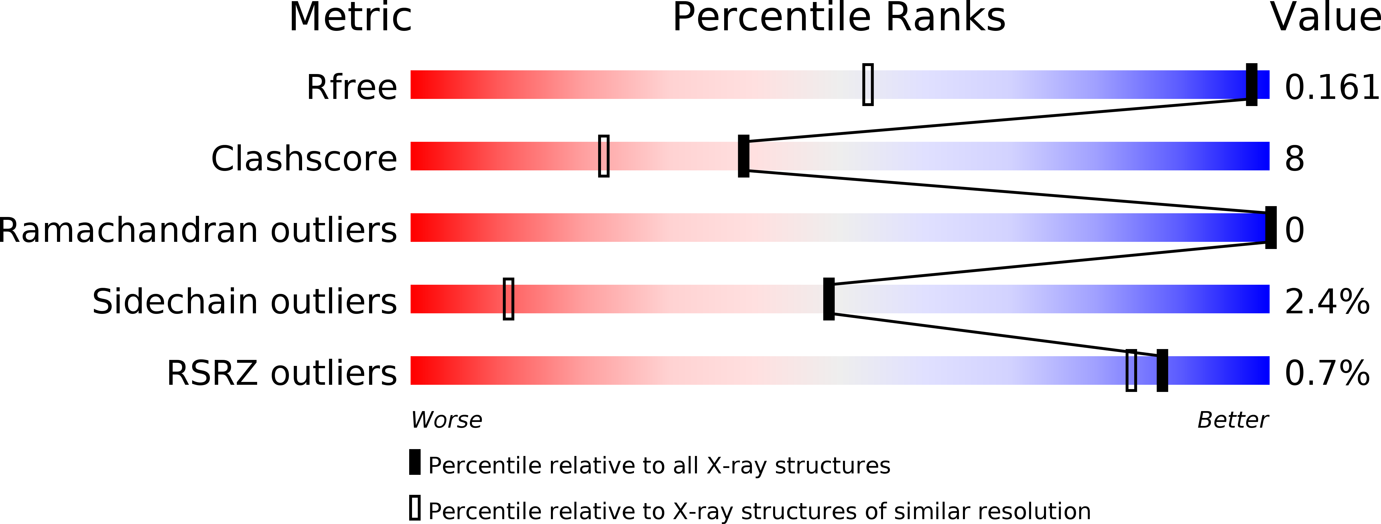
R-Value Free:
0.18
R-Value Observed:
0.15
Space Group:
P 21 21 21