3ZSE image
Deposition Date 2011-06-27
Release Date 2012-02-08
Last Version Date 2024-10-23
Entry Detail
PDB ID:
3ZSE
Keywords:
Title:
3D Structure of a thermophilic family GH11 xylanase from Thermobifida fusca
Biological Source:
Source Organism:
Host Organism:
Method Details:
Experimental Method:
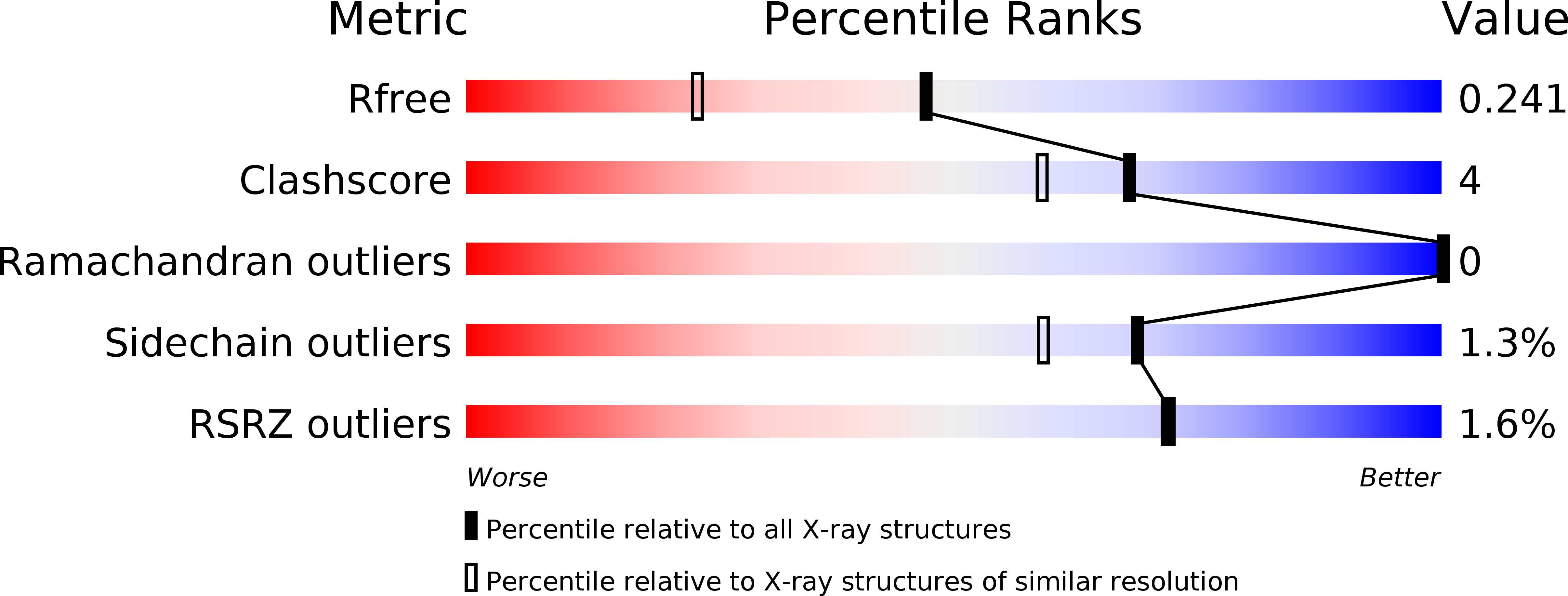
Resolution:
1.78 Å
R-Value Free:
0.24
R-Value Work:
0.19
R-Value Observed:
0.19
Space Group:
P 21 21 21
Macromolecular Entities
Structures with similar UniProt ID
Protein Blast
Polymer Type:polypeptide(L)
Molecule:ENDO-1,4-BETA-XYLANASE
Gene (Uniprot):xyl11
Chain IDs:A
Chain Length:201
Number of Molecules:1
Biological Source:THERMOBIFIDA FUSCA
Ligand Molecules
Primary Citation
Three-Dimensional Structure of a Thermophilic Family Gh11 Xylanase from Thermobifida Fusca.
Acta Crystallogr.,Sect.F 68 141 ? (2012)
PMID: 22297985 DOI: 10.1107/S1744309111049608

Abstact

Thermostable enzymes employ various structural features dictated at the amino-acid sequence level that allow them to maintain their integrity at higher temperatures. Many hypotheses as to the nature of thermal stability have been proposed, including optimized core hydrophobicity and an increase in charged surface residues to enhance polar solvent interactions for solubility. Here, the three-dimensional structure of the family GH11 xylanase from the moderate thermophile Thermobifida fusca in its trapped covalent glycosyl-enzyme intermediate complex is presented. Interactions with the bound ligand show fewer direct hydrogen bonds from ligand to protein than observed in previous complexes from other species and imply that binding of the xylan substrate involves several water-mediated hydrogen bonds.

Legend

Protein

Chemical

Disease

Primary Citation of related structures