3ZRS image
Deposition Date 2011-06-17
Release Date 2012-01-11
Last Version Date 2023-12-20
Entry Detail
PDB ID:
3ZRS
Keywords:
Title:
X-ray crystal structure of a KirBac potassium channel highlights a mechanism of channel opening at the bundle-crossing gate.
Biological Source:
Source Organism:
Host Organism:
Method Details:
Experimental Method:
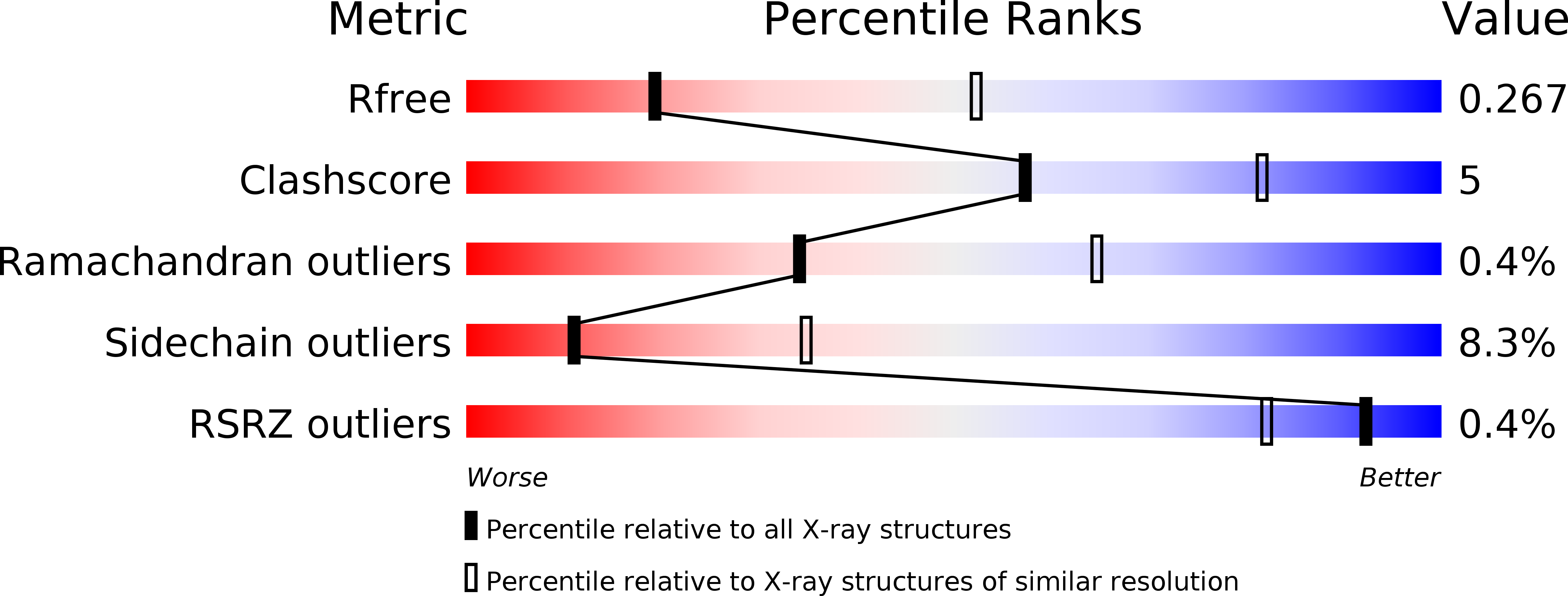
Resolution:
3.05 Å
R-Value Free:
0.25
R-Value Work:
0.22
R-Value Observed:
0.22
Space Group:
P 4 21 2
Macromolecular Entities
Structures with similar UniProt ID
Protein Blast
Polymer Type:polypeptide(L)
Molecule:ATP-SENSITIVE INWARD RECTIFIER POTASSIUM CHANNEL 10
Mutagens:YES
Chain IDs:A
Chain Length:301
Number of Molecules:1
Biological Source:MAGNETOSPIRILLUM MAGNETOTACTICUM
Primary Citation
Structure of a Kirbac Potassium Channel with an Open Bundle Crossing Indicates a Mechanism of Channel Gating
Nat.Struct.Mol.Biol. 19 158 ? (2012)
PMID: 22231399 DOI: 10.1038/NSMB.2208

Abstact

KirBac channels are prokaryotic homologs of mammalian inwardly rectifying (Kir) potassium channels, and recent crystal structures of both Kir and KirBac channels have provided major insight into their unique structural architecture. However, all of the available structures are closed at the helix bundle crossing, and therefore the structural mechanisms that control opening of their primary activation gate remain unknown. In this study, we engineered the inner pore-lining helix (TM2) of KirBac3.1 to trap the bundle crossing in an apparently open conformation and determined the crystal structure of this mutant channel to 3.05 Å resolution. Contrary to previous speculation, this new structure suggests a mechanistic model in which rotational 'twist' of the cytoplasmic domain is coupled to opening of the bundle-crossing gate through a network of inter- and intrasubunit interactions that involve the TM2 C-linker, slide helix, G-loop and the CD loop.

Legend

Protein

Chemical

Disease

Primary Citation of related structures