
Deposition Date
2013-02-25
Release Date
2013-08-14
Last Version Date
2024-10-09
Entry Detail
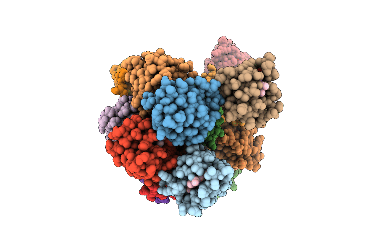
PDB ID:
3ZOW
Keywords:
Title:
Crystal Structure of Wild Type Nitrosomonas europaea Cytochrome c552
Biological Source:
Source Organism(s):
NITROSOMONAS EUROPAEA (Taxon ID: 915)
Method Details:
Experimental Method:
Resolution:
2.35 Å
R-Value Free:
0.25
R-Value Work:
0.20
R-Value Observed:
0.20
Space Group:
P 21 21 21
