
Deposition Date
2013-02-22
Release Date
2013-08-21
Last Version Date
2023-12-20
Entry Detail
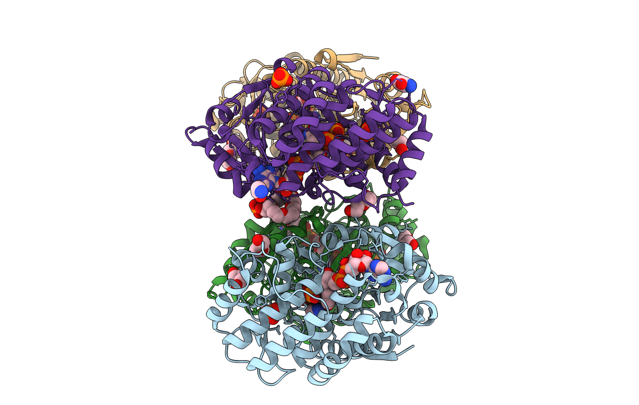
PDB ID:
3ZOK
Keywords:
Title:
Structure of 3-Dehydroquinate Synthase from Actinidia chinensis in complex with NAD
Biological Source:
Source Organism(s):
ACTINIDIA CHINENSIS (Taxon ID: 3625)
Expression System(s):
Method Details:
Experimental Method:
Resolution:
2.40 Å
R-Value Free:
0.25
R-Value Work:
0.18
R-Value Observed:
0.18
Space Group:
P 1 21 1
