3ZHO image
Deposition Date 2012-12-23
Release Date 2013-08-28
Last Version Date 2023-12-20
Entry Detail
PDB ID:
3ZHO
Keywords:
Title:
X-ray structure of E.coli Wrba in complex with FMN at 1.2 A resolution
Biological Source:
Source Organism:
ESCHERICHIA COLI (Taxon ID: 562)
Host Organism:
Method Details:
Experimental Method:
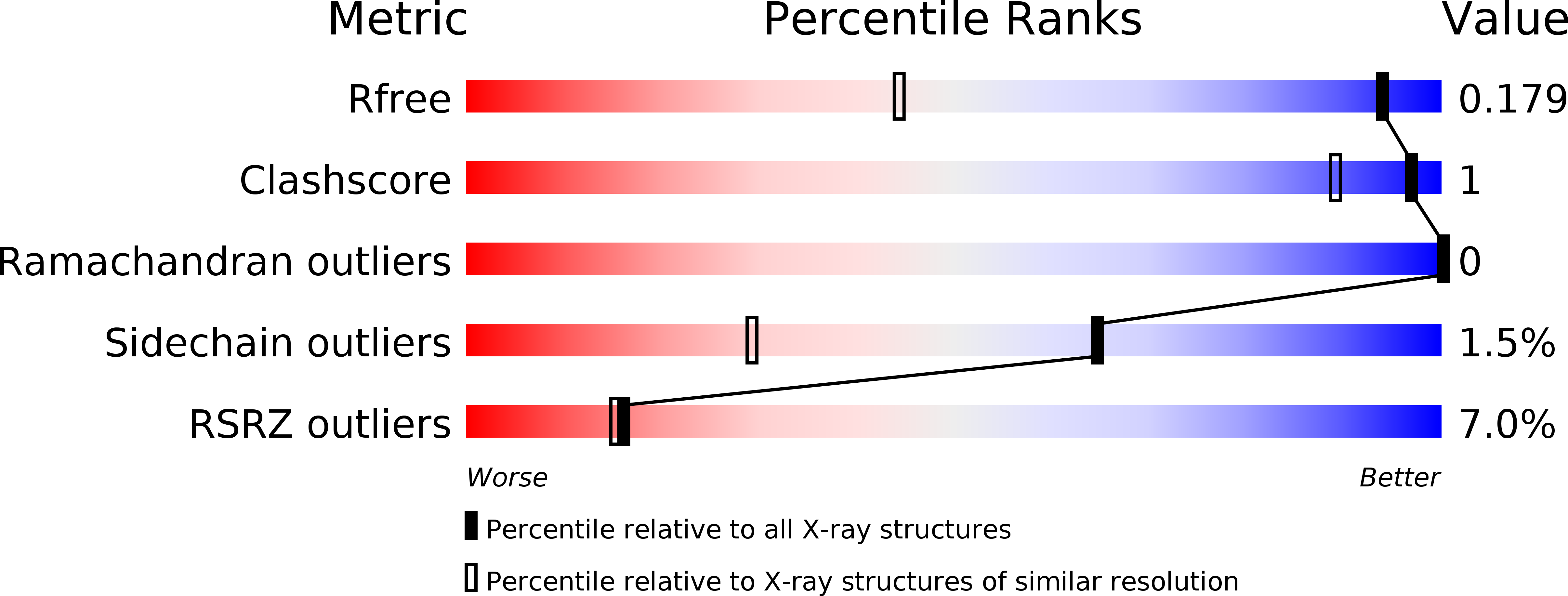
Resolution:
1.20 Å
R-Value Free:
0.17
R-Value Work:
0.14
R-Value Observed:
0.14
Space Group:
P 41 21 2
Macromolecular Entities
Structures with similar UniProt ID
Protein Blast
Polymer Type:polypeptide(L)
Molecule:FLAVOPROTEIN WRBA
Gene (Uniprot):wrbA
Chain IDs:A, B
Chain Length:197
Number of Molecules:2
Biological Source:ESCHERICHIA COLI
Modified Residue
Compound ID Chain ID Parent Comp ID Details 2D Image
SME A MET METHIONINE SULFOXIDE
Ligand Molecules
Primary Citation
1.2 A Resolution Crystal Structure of Escherichia Coli Wrba Holoprotein
Acta Crystallogr.,Sect.D 69 1757 ? (2013)
PMID: 23999298 DOI: 10.1107/S0907444913017162

Abstact

The Escherichia coli protein WrbA, an FMN-dependent NAD(P)H:quinone oxidoreductase, was crystallized under new conditions in the presence of FAD or the native cofactor FMN. Slow-growing deep yellow crystals formed with FAD display the tetragonal bipyramidal shape typical for WrbA and diffract to 1.2 Å resolution, the highest yet reported. Faster-growing deep yellow crystals formed with FMN display an atypical shape, but diffract to only ∼1.6 Å resolution and are not analysed further here. The 1.2 Å resolution structure detailed here revealed only FMN in the active site and no electron density that can accommodate the missing parts of FAD. The very high resolution supports the modelling of the FMN isoalloxazine with a small but distinct propeller twist, apparently the first experimental observation of this predicted conformation, which appears to be enforced by the protein through a network of hydrogen bonds. Comparison of the electron density of the twisted isoalloxazine ring with the results of QM/MM simulations is compatible with the oxidized redox state. The very high resolution also supports the unique refinement of Met10 as the sulfoxide, confirmed by mass spectrometry. Bond lengths, intramolecular distances, and the pattern of hydrogen-bond donors and acceptors suggest the cofactor may interact with Met10. Slow incorporation of FMN, which is present as a trace contaminant in stocks of FAD, into growing crystals may be responsible for the near-atomic resolution, but a direct effect of the conformation of FMN and/or Met10 sulfoxide cannot be ruled out.

Legend

Protein

Chemical

Disease

Primary Citation of related structures