3ZE3 image
Entry Detail
PDB ID:
3ZE3
Keywords:
Title:
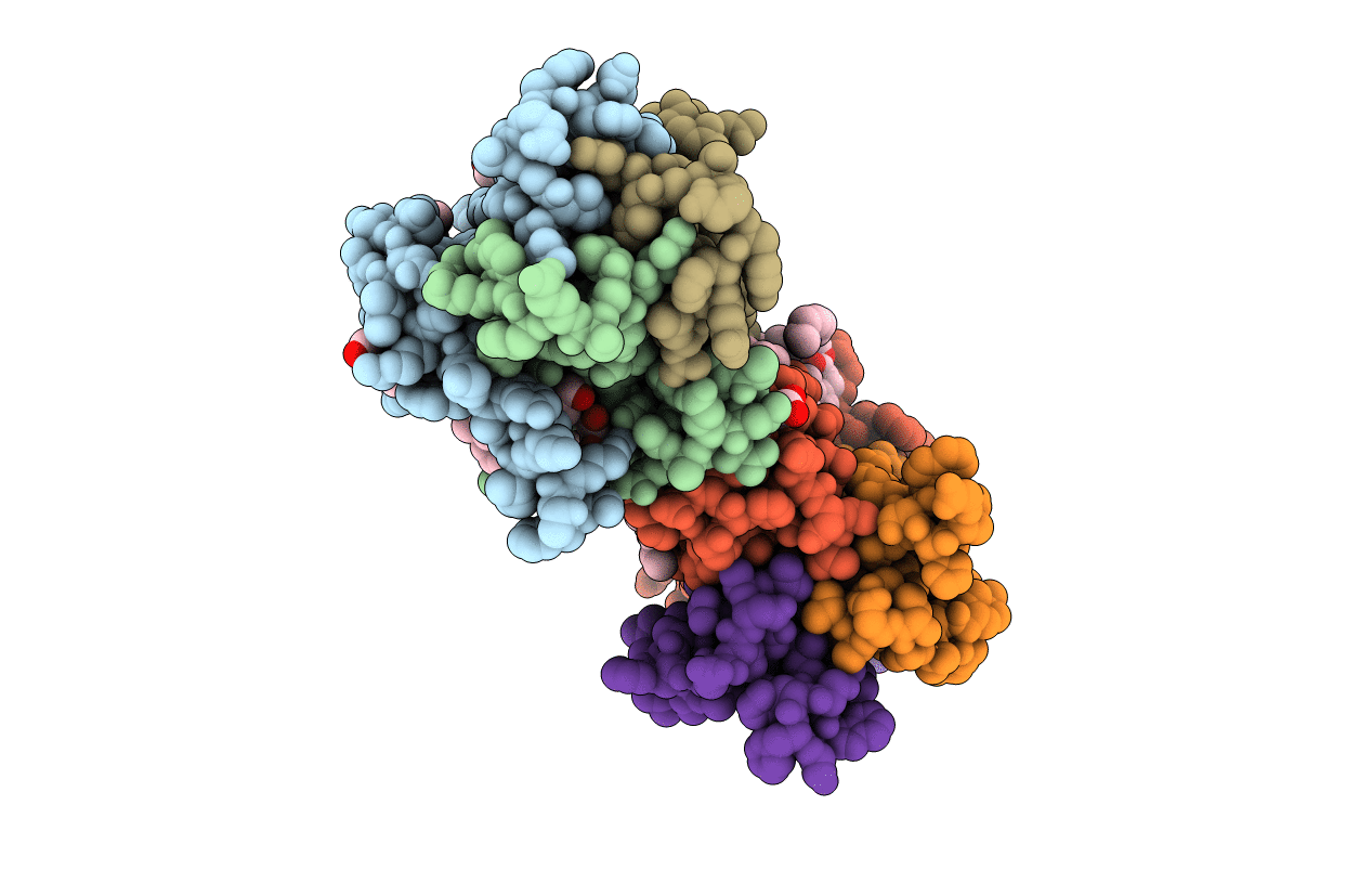
Crystal structure of the integral membrane diacylglycerol kinase - delta7
Biological Source:
Source Organism:
Host Organism:
PDB Version:
Deposition Date:
2012-12-03
Release Date:
2013-05-22
Method Details:
Experimental Method:
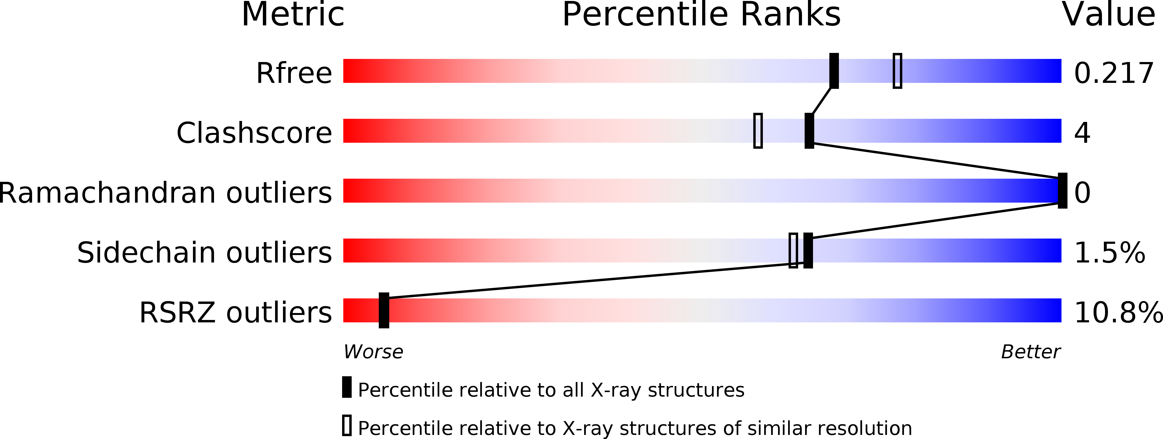
Resolution:
2.05 Å
R-Value Free:
0.21
R-Value Work:
0.19
R-Value Observed:
0.19
Space Group:
P 21 21 21
Macromolecular Entities
Polymer Type:polypeptide(L)
Description:DIACYLGLYCEROL KINASE
Mutations:YES
Chain IDs:A, B, C, D, E, F
Chain Length:130
Number of Molecules:6
Biological Source:ESCHERICHIA COLI K-12
Primary Citation
Crystal Structure of the Integral Membrane Diacylglycerol Kinase.
Nature 497 521 ? (2013)
PMID: 23676677 DOI: 10.1038/NATURE12179

Abstact

Diacylglycerol kinase catalyses the ATP-dependent phosphorylation of diacylglycerol to phosphatidic acid for use in shuttling water-soluble components to membrane-derived oligosaccharide and lipopolysaccharide in the cell envelope of Gram-negative bacteria. For half a century, this 121-residue kinase has served as a model for investigating membrane protein enzymology, folding, assembly and stability. Here we present crystal structures for three functional forms of this unique and paradigmatic kinase, one of which is wild type. These reveal a homo-trimeric enzyme with three transmembrane helices and an amino-terminal amphiphilic helix per monomer. Bound lipid substrate and docked ATP identify the putative active site that is of the composite, shared site type. The crystal structures rationalize extensive biochemical and biophysical data on the enzyme. They are, however, at variance with a published solution NMR model in that domain swapping, a key feature of the solution form, is not observed in the crystal structures.

Legend

Protein

Chemical

Disease

Primary Citation of related structures