3ZCJ image
Deposition Date 2012-11-20
Release Date 2013-10-09
Last Version Date 2023-12-20
Entry Detail
PDB ID:
3ZCJ
Keywords:
Title:
Crystal structure of Helicobacter pylori T4SS protein CagL in a tetragonal crystal form with a helical RGD-motif (6 Mol per ASU)
Biological Source:
Source Organism:
Host Organism:
Method Details:
Experimental Method:
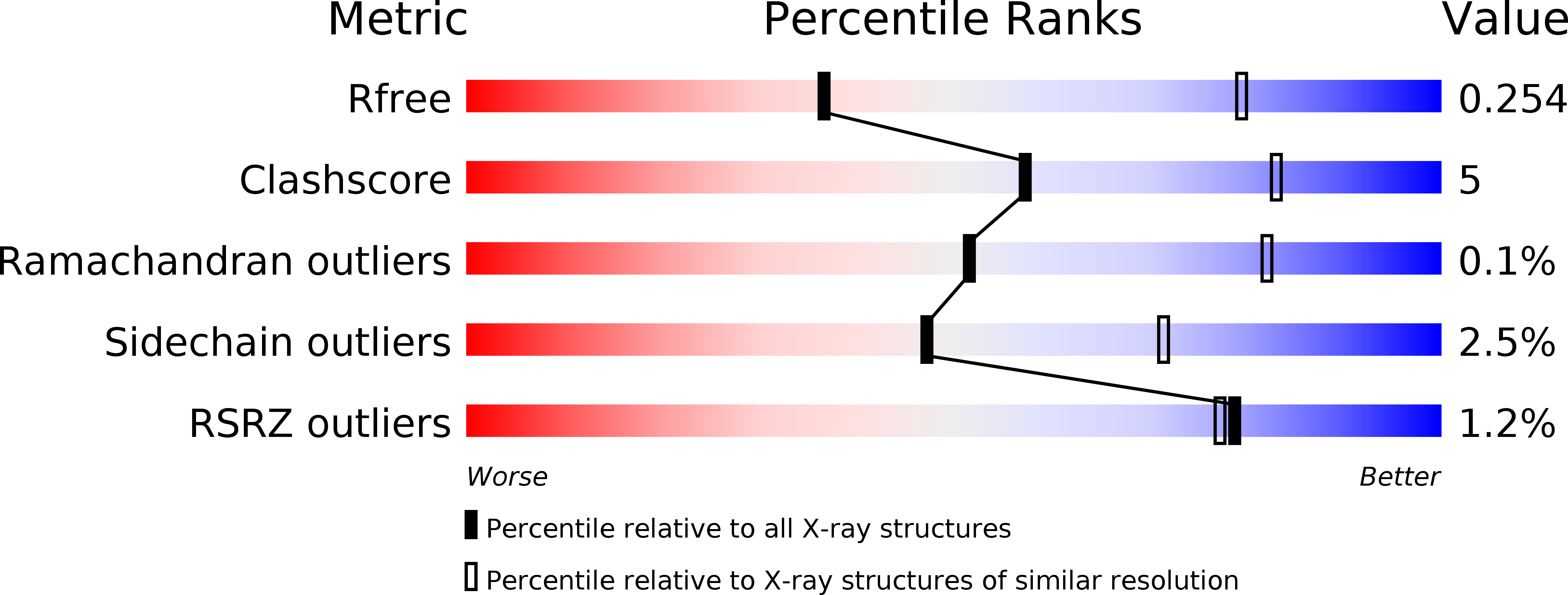
Resolution:
3.25 Å
R-Value Free:
0.25
R-Value Work:
0.20
R-Value Observed:
0.20
Space Group:
P 43 21 2
Macromolecular Entities
Structures with similar UniProt ID
Protein Blast
Polymer Type:polypeptide(L)
Molecule:CAGL
Gene (Uniprot):HP_0539
Chain IDs:A, B, C, D, E, F
Chain Length:220
Number of Molecules:6
Biological Source:HELICOBACTER PYLORI
Modified Residue
Compound ID Chain ID Parent Comp ID Details 2D Image
MLY A LYS N-DIMETHYL-LYSINE
Primary Citation
A Helical Rgd Motif Promoting Cell Adhesion: Crystal Structures of the Helicobacter Pylori Type Iv Secretion System Pilus Protein Cagl
Structure 21 1931 ? (2013)
PMID: 24076404 DOI: 10.1016/J.STR.2013.08.018

Abstact

RGD tripeptide motifs frequently mediate ligand binding to integrins. The type IV secretion system (T4SS) protein CagL of the gastric pathogen Helicobacter pylori also contains an RGD motif. CagL decorates the T4SS pilus and may function as an adhesin for host cells. Whether CagL binds integrins via its RGD motif is under debate. Here, we present crystal structures of CagL revealing an elongated four-helix bundle that appears evolutionarily unrelated to the proposed VirB5 orthologs. The RGD motif is surface-exposed but located within a long α helix. This is unprecedented as previously characterized integrin-binding RGD motifs are located within extended or flexible loops. Yet, adhesion of gastric epithelial cells to CagL was strictly RGD-dependent. Comparison of seven crystallographically independent molecules reveals substantial structural flexibility. Intramolecular disulfide bonds engineered to reduce CagL flexibility resulted in more stable protein, but unable to support cell adhesion. CagL may thus partly unfold during receptor binding.

Legend

Protein

Chemical

Disease

Primary Citation of related structures