3ZCB image
Deposition Date 2012-11-19
Release Date 2013-06-19
Last Version Date 2023-12-20
Entry Detail
PDB ID:
3ZCB
Title:
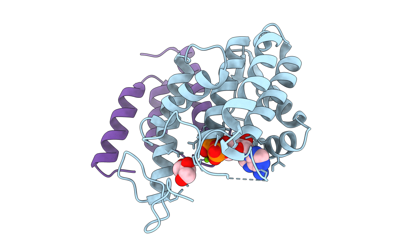
VbhT Fic protein from Bartonella schoenbuchensis in complex with VbhA antitoxin mutant E24G and ATP
Biological Source:
Source Organism:
Host Organism:
Method Details:
Experimental Method:
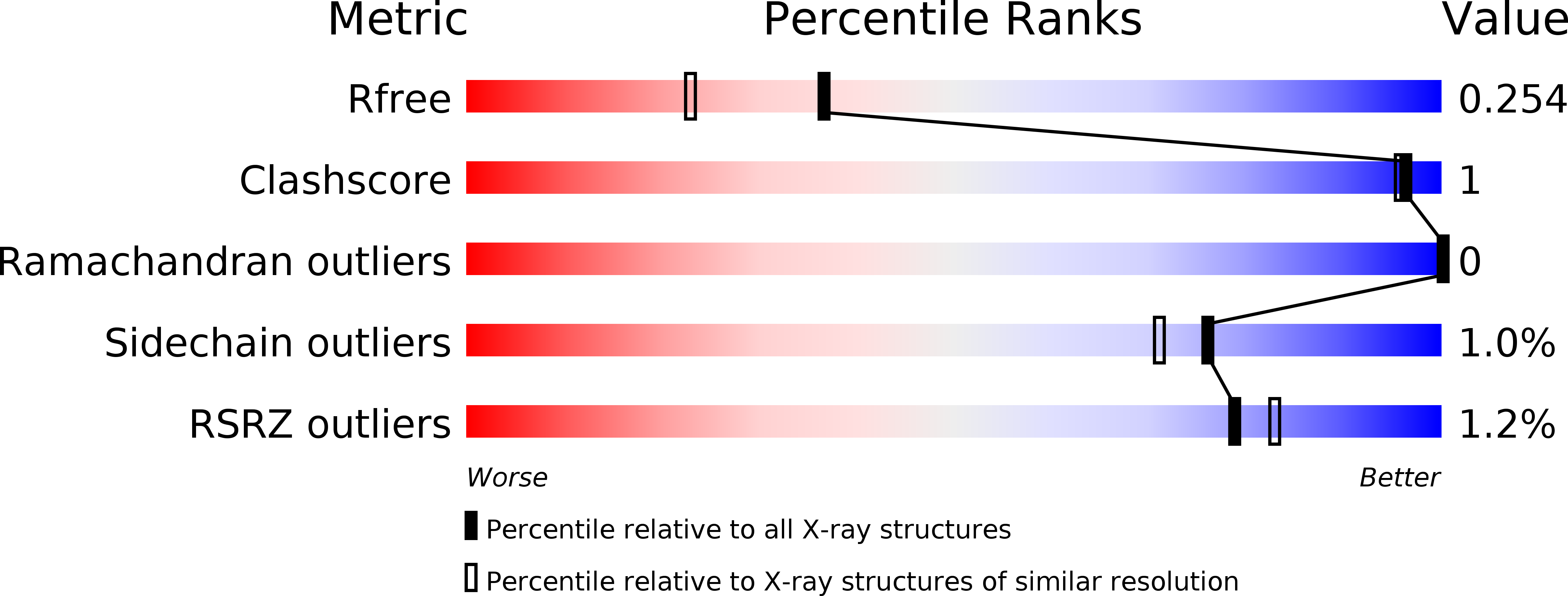
Resolution:
1.94 Å
R-Value Free:
0.24
R-Value Work:
0.19
R-Value Observed:
0.19
Space Group:
C 1 2 1
Macromolecular Entities
Structures with similar UniProt ID
Protein Blast
Polymer Type:polypeptide(L)
Molecule:ADENOSINE MONOPHOSPHATE-PROTEIN TRANSFERASE VBHT
Gene (Uniprot):vbhT
Chain IDs:A
Chain Length:198
Number of Molecules:1
Biological Source:BARTONELLA SCHOENBUCHENSIS
Structures with similar UniProt ID
Protein Blast
Polymer Type:polypeptide(L)
Molecule:ANTITOXIN VBHA
Gene (Uniprot):B11C_100027
Mutagens:YES
Chain IDs:B
Chain Length:62
Number of Molecules:1
Biological Source:BARTONELLA SCHOENBUCHENSIS
Primary Citation
Conserved Inhibitory Mechanism and Competent ATP Binding Mode for Adenylyltransferases with Fic Fold.
Plos One 8 64901 ? (2013)
PMID: 23738009 DOI: 10.1371/JOURNAL.PONE.0064901

Abstact

The ubiquitous FIC domain is evolutionarily conserved from bacteria to human and has been shown to catalyze AMP transfer onto protein side-chain hydroxyl groups. Recently, it was predicted that most catalytically competent Fic proteins are inhibited by the presence of an inhibitory helix αinh that is provided by a cognate anti-toxin (class I), or is part of the N- or C-terminal part of the Fic protein itself (classes II and III). In vitro, inhibition is relieved by mutation of a conserved glutamate of αinh to glycine. For the class III bacterial Fic protein NmFic from Neisseria meningitidis, the inhibitory mechanism has been elucidated. Here, we extend above study by including bacterial class I and II Fic proteins VbhT from Bartonella schoenbuchensis and SoFic from Shewanella oneidensis, respectively, and the respective E->G mutants. Comparative enzymatic and crystallographic analyses show that, in all three classes, the ATP substrate binds to the wild-type FIC domains, but with the α-phosphate in disparate and non-competent orientations. In the E->G mutants, however, the tri-phosphate moiety is found reorganized to the same tightly bound structure through a unique set of hydrogen bonds with Fic signature motif residues. The γ-phosphate adopts the location that is taken by the inhibitory glutamate in wild-type resulting in an α-phosphate orientation that can be attacked in-line by a target side-chain hydroxyl group. The latter is properly registered to the Fic active center by main-chain β-interactions with the β-hairpin flap. These data indicate that the active site motif and the exposed edge of the flap are both required to form an adenylylation-competent Fic protein.

Legend

Protein

Chemical

Disease

Primary Citation of related structures