Deposition Date
2015-01-02
Release Date
2015-12-16
Last Version Date
2024-11-13
Entry Detail
PDB ID:
3X2U
Keywords:
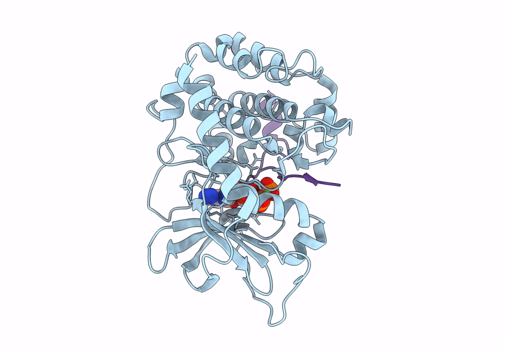
Title:
Michaelis-like initial complex of cAMP-dependent Protein Kinase Catalytic Subunit.
Biological Source:
Source Organism(s):
Mus musculus (Taxon ID: 10090)
synthetic construct (Taxon ID: 32630)
synthetic construct (Taxon ID: 32630)
Expression System(s):
Method Details:
Experimental Method:
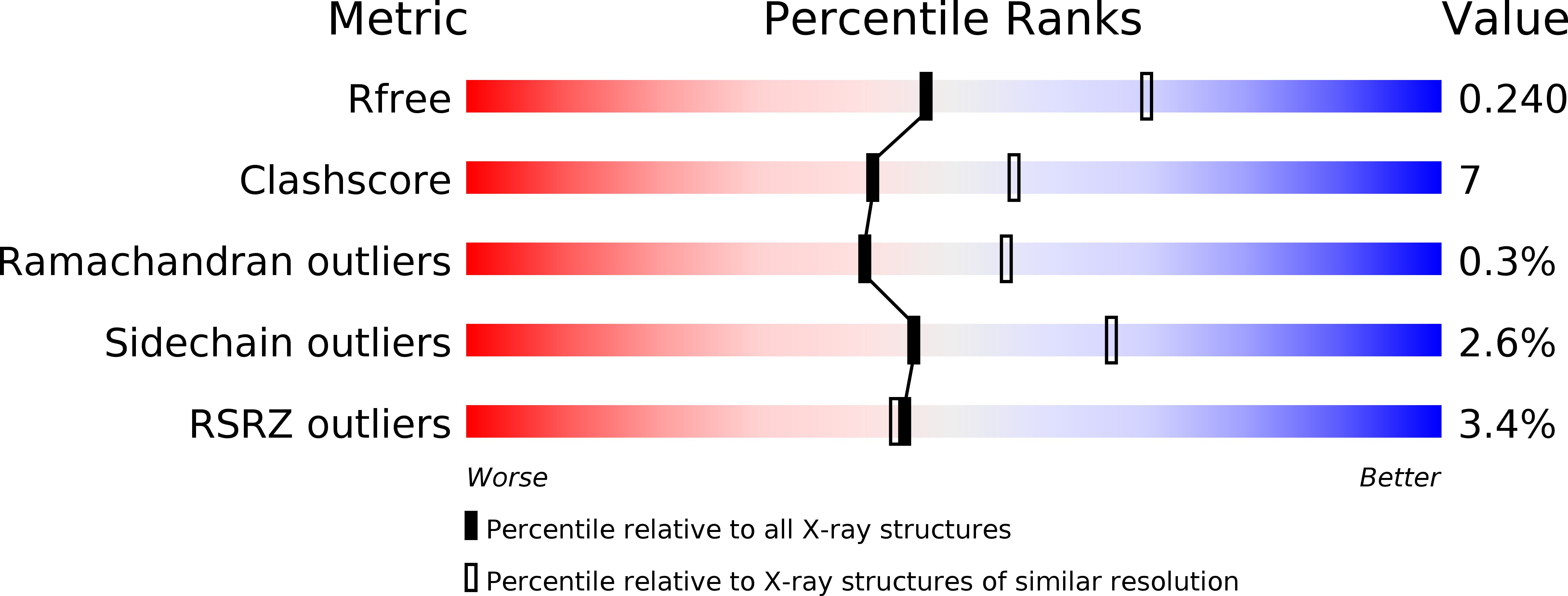
Resolution:
2.40 Å
R-Value Free:
0.23
R-Value Work:
0.20
R-Value Observed:
0.20
Space Group:
P 21 21 21