
Deposition Date
2014-12-22
Release Date
2015-10-07
Last Version Date
2025-10-29
Entry Detail
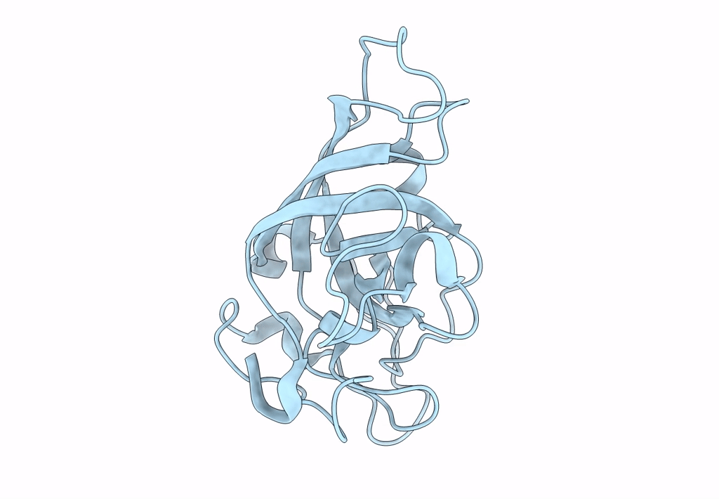
PDB ID:
3X2O
Keywords:
Title:
Neutron and X-ray joint refined structure of PcCel45A apo form at 298K.
Biological Source:
Source Organism(s):
Phanerochaete chrysosporium (Taxon ID: 5306)
Expression System(s):
Method Details:
Experimental Method:
R-Value Free:
['0.25
R-Value Work:
['0.22
R-Value Observed:
['0.22
Space Group:
P 21 21 21
