3X29 image
Deposition Date 2014-12-13
Release Date 2015-01-21
Last Version Date 2024-10-16
Entry Detail
PDB ID:
3X29
Keywords:
Title:
CRYSTAL STRUCTURE of MOUSE CLAUDIN-19 IN COMPLEX with C-TERMINAL FRAGMENT OF CLOSTRIDIUM PERFRINGENS ENTEROTOXIN
Biological Source:
Source Organism:
Host Organism:
Method Details:
Experimental Method:
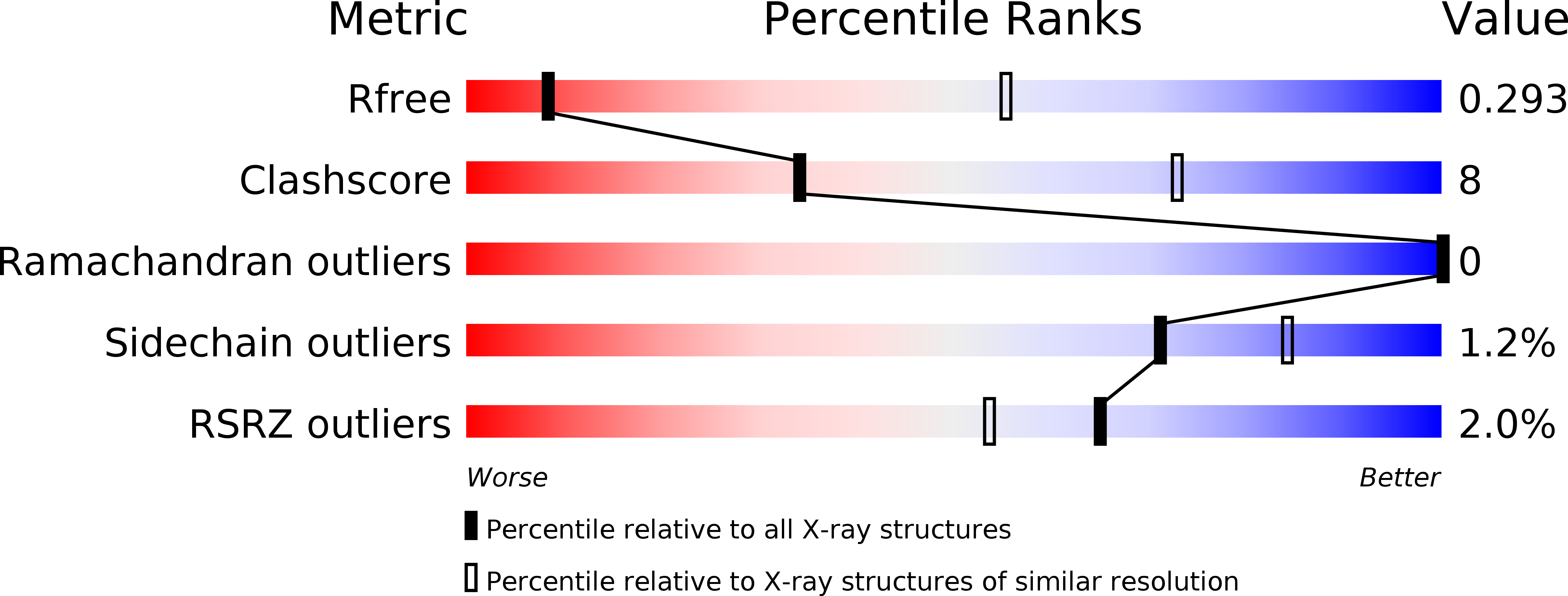
Resolution:
3.70 Å
R-Value Free:
0.29
R-Value Work:
0.25
R-Value Observed:
0.25
Space Group:
P 21 21 21
Macromolecular Entities
Structures with similar UniProt ID
Protein Blast
Polymer Type:polypeptide(L)
Molecule:Claudin-19
Gene (Uniprot):Cldn19
Mutagens:C104A, C183A, C184A
Chain IDs:A, C
Chain Length:185
Number of Molecules:2
Biological Source:Mus musculus
Structures with similar UniProt ID
Protein Blast
Polymer Type:polypeptide(L)
Molecule:Heat-labile enterotoxin B chain
Gene (Uniprot):cpe
Mutagens:S313A
Chain IDs:B, D
Chain Length:119
Number of Molecules:2
Biological Source:Clostridium perfringens
Ligand Molecules
Primary Citation
Structural insight into tight junction disassembly by Clostridium perfringens enterotoxin
Science 347 775 778 (2015)
PMID: 25678664 DOI: 10.1126/science.1261833

Abstact

The C-terminal region of Clostridium perfringens enterotoxin (C-CPE) can bind to specific claudins, resulting in the disintegration of tight junctions (TJs) and an increase in the paracellular permeability across epithelial cell sheets. Here we present the structure of mammalian claudin-19 in complex with C-CPE at 3.7 Å resolution. The structure shows that C-CPE forms extensive hydrophobic and hydrophilic interactions with the two extracellular segments of claudin-19. The claudin-19/C-CPE complex shows no density of a short extracellular helix that is critical for claudins to assemble into TJ strands. The helix displacement may thus underlie C-CPE-mediated disassembly of TJs.

Legend

Protein

Chemical

Disease

Primary Citation of related structures