Deposition Date
2014-10-23
Release Date
2015-02-25
Last Version Date
2024-05-29
Entry Detail
PDB ID:
3X0Y
Keywords:
Title:
Crystal structure of FMN-bound DszC from Rhodococcus erythropolis D-1
Biological Source:
Source Organism(s):
Rhodococcus erythropolis (Taxon ID: 1833)
Expression System(s):
Method Details:
Experimental Method:
Resolution:
2.30 Å
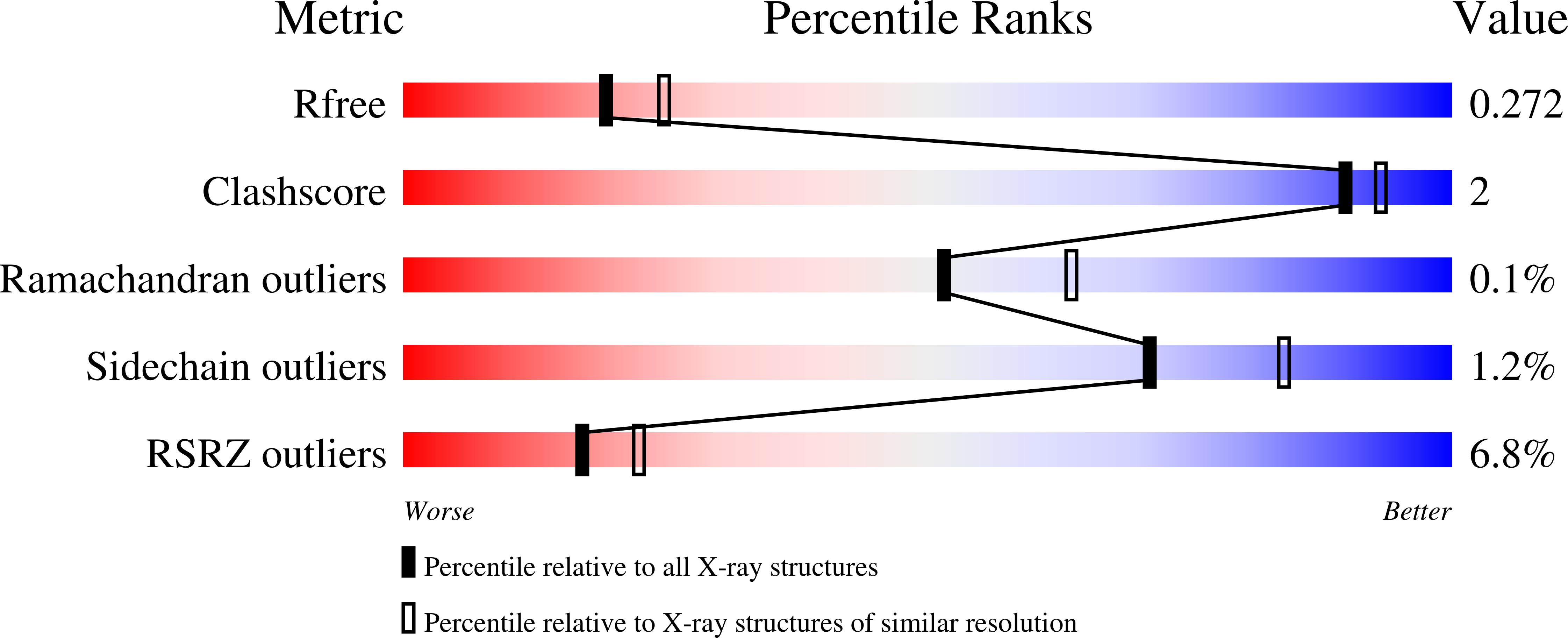
R-Value Free:
0.27
R-Value Work:
0.22
R-Value Observed:
0.23
Space Group:
C 1 2 1