3WZS image
Entry Detail
PDB ID:
3WZS
Keywords:
Title:
Crystal structure of Trx3 domain of UGGT (detergent-bound form)
Biological Source:
PDB Version:
Deposition Date:
2014-10-03
Release Date:
2014-12-03
Method Details:
Experimental Method:
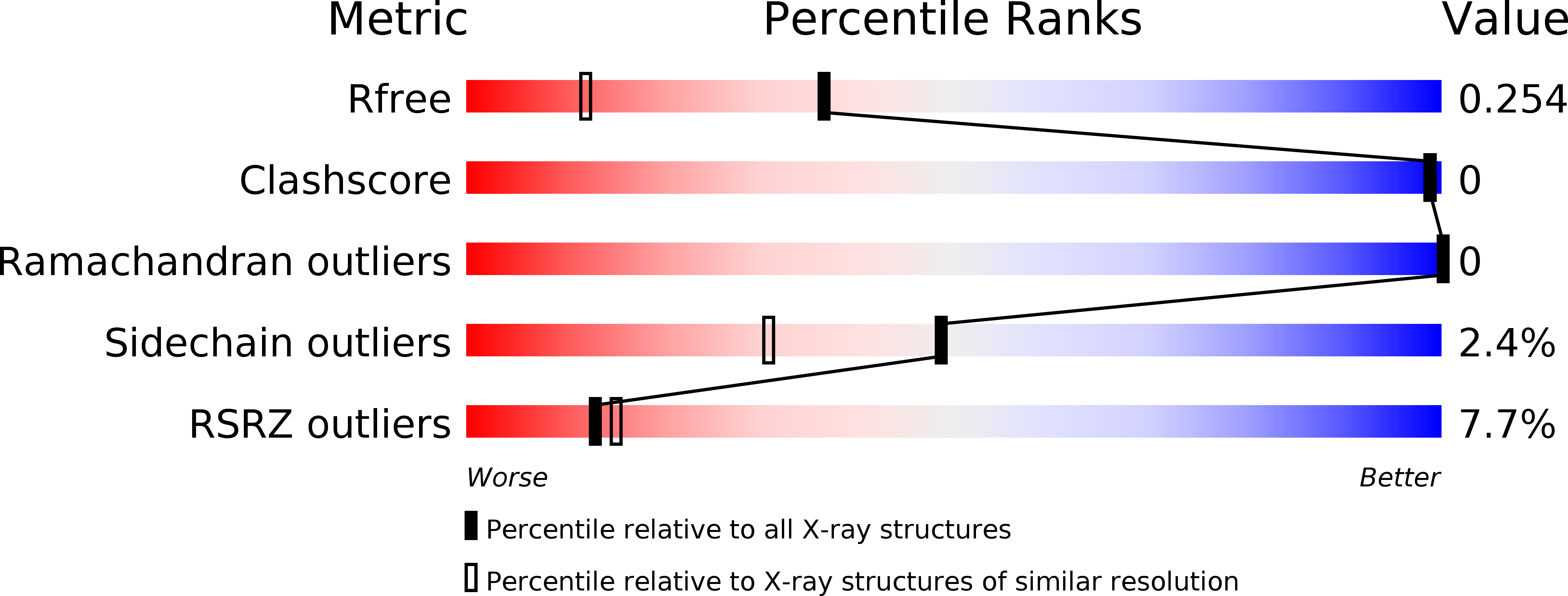
Resolution:
1.70 Å
R-Value Free:
0.24
R-Value Work:
0.20
R-Value Observed:
0.20
Space Group:
C 2 2 21
Macromolecular Entities
Polymer Type:polypeptide(L)
Description:UDP-glucose-glycoprotein glucosyltransferase-like protein
Chain IDs:A
Chain Length:163
Number of Molecules:1
Biological Source:Chaetomium thermophilum var. thermophilum DSM 1495
Modified Residue
Compound ID Chain ID Parent Comp ID Details 2D Image
MSE A MET SELENOMETHIONINE
Ligand Molecules
Primary Citation
Structural insight into substrate recognition by the endoplasmic reticulum folding-sensor enzyme: crystal structure of third thioredoxin-like domain of UDP-glucose:glycoprotein glucosyltransferase
Sci Rep 4 7322 7322 (2014)
PMID: 25471383 DOI: 10.1038/srep07322

Abstact

The endoplasmic reticulum (ER) possesses a protein quality control system that supports the efficient folding of newly synthesized glycoproteins. In this system, a series of N-linked glycan intermediates displayed on proteins serve as quality tags. The ER folding-sensor enzyme UDP-glucose:glycoprotein glucosyltransferase (UGGT) operates as the gatekeeper for ER quality control by specifically transferring monoglucose residues to incompletely folded glycoproteins, thereby allowing them to interact with lectin chaperone complexes to facilitate their folding. Despite its functional importance, no structural information is available for this key enzyme to date. To elucidate the folding-sensor mechanism in the ER, we performed a structural study of UGGT. Based on bioinformatics analyses, the folding-sensor region of UGGT was predicted to harbour three tandem thioredoxin (Trx)-like domains, which are often found in proteins involved in ER quality control. Furthermore, we determined the three-dimensional structure of the third Trx-like domain, which exhibits an extensive hydrophobic patch concealed by its flexible C-terminal helix. Our structural data suggest that this hydrophobic patch is involved in intermolecular interactions, thereby contributing to the folding-sensor mechanism of UGGT.

Legend

Protein

Chemical

Disease

Primary Citation of related structures