3WZ2 image
Deposition Date 2014-09-18
Release Date 2014-10-08
Last Version Date 2024-03-20
Entry Detail
PDB ID:
3WZ2
Keywords:
Title:
Crystal structure of Pyrococcus furiosus PbaA, an archaeal homolog of proteasome-assembly chaperone
Biological Source:
Source Organism:
Method Details:
Experimental Method:
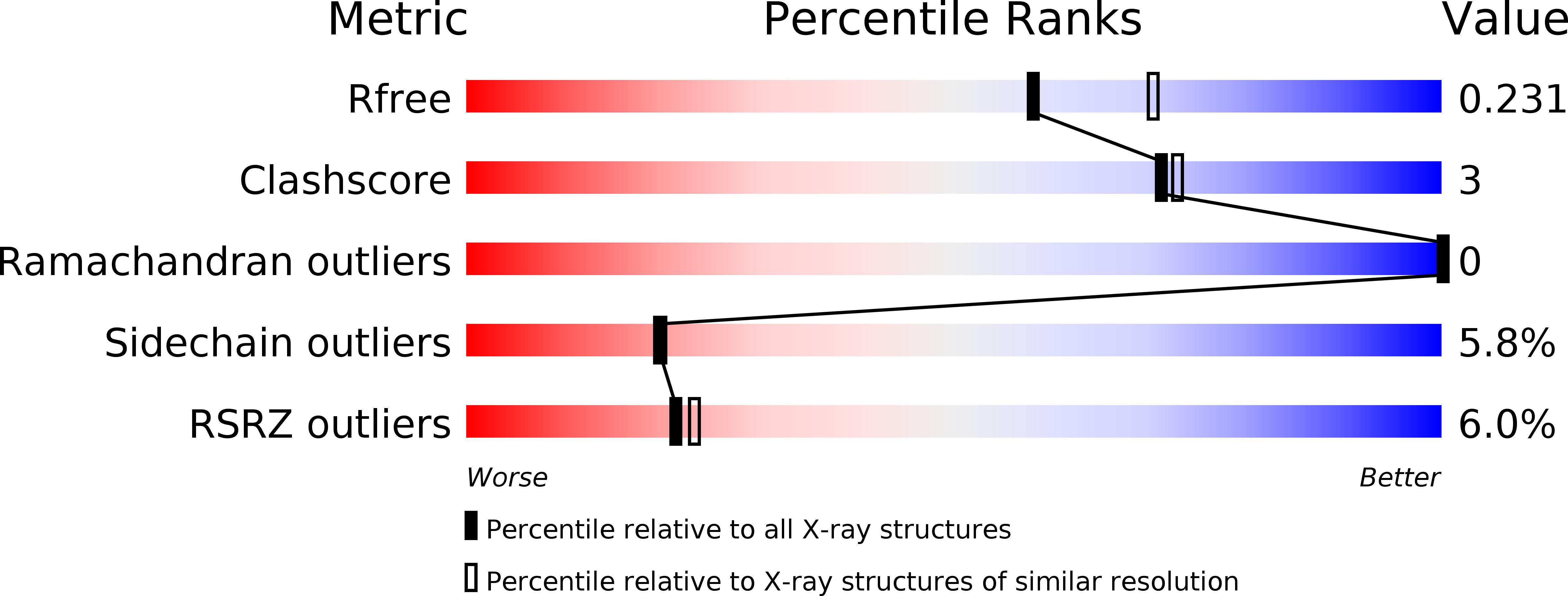
Resolution:
2.25 Å
R-Value Free:
0.22
R-Value Work:
0.19
R-Value Observed:
0.19
Space Group:
C 2 2 21
Macromolecular Entities
Structures with similar UniProt ID
Protein Blast
Polymer Type:polypeptide(L)
Molecule:Uncharacterized protein
Gene (Uniprot):PF0015
Chain IDs:A, B, C, D, E
Chain Length:245
Number of Molecules:5
Biological Source:Pyrococcus furiosus DSM 3638
Primary Citation
Crystal structure of archaeal homolog of proteasome-assembly chaperone PbaA
Biochem.Biophys.Res.Commun. 453 493 497 (2014)
PMID: 25285636 DOI: 10.1016/j.bbrc.2014.09.114

Abstact

Formation of the eukaryotic proteasome is not a spontaneous process but a highly ordered process assisted by several assembly chaperones. In contrast, archaeal proteasome subunits can spontaneously assemble into an active form. Recent bioinformatic analysis identified the proteasome-assembly chaperone-like proteins, PbaA and PbaB, in archaea. Our previous study showed that the PbaB homotetramer functions as a proteasome activator through its tentacle-like C-terminal segments. However, a functional role of the other homolog PbaA has remained elusive. Here we determined the 2.25-Å resolution structure of PbaA, illustrating its disparate tertiary and quaternary structures compared with PbaB. PbaA forms a homopentamer in which the C-terminal segments, with a putative proteasome-activating motif, are packed against the core. These findings offer deeper insights into the molecular evolution relationships between the proteasome-assembly chaperones and the proteasome activators.

Legend

Protein

Chemical

Disease

Primary Citation of related structures
Feedback Form
Name
Email
Institute
Feedback