3WYC image
Deposition Date 2014-08-25
Release Date 2015-05-06
Last Version Date 2024-03-20
Entry Detail
PDB ID:
3WYC
Keywords:
Title:
Structure of a meso-diaminopimelate dehydrogenase in complex with NADP
Biological Source:
Source Organism:
Host Organism:
Method Details:
Experimental Method:
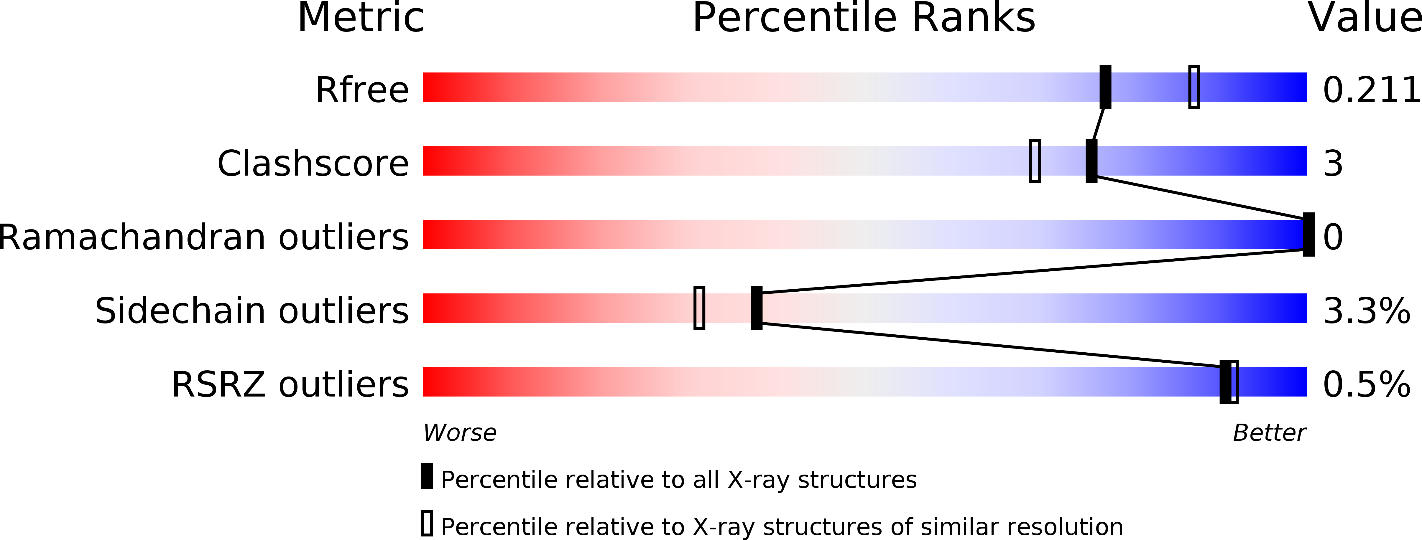
Resolution:
2.07 Å
R-Value Free:
0.20
R-Value Work:
0.17
R-Value Observed:
0.17
Space Group:
H 3
Macromolecular Entities
Structures with similar UniProt ID
Protein Blast
Polymer Type:polypeptide(L)
Molecule:Meso-diaminopimelate D-dehydrogenase
Gene (Uniprot):ddh
Chain IDs:A, B
Chain Length:334
Number of Molecules:2
Biological Source:Ureibacillus thermosphaericus
Primary Citation
Structural insight into the thermostable NADP(+)-dependent meso-diaminopimelate dehydrogenase from Ureibacillus thermosphaericus
Acta Crystallogr.,Sect.D 71 1136 1146 (2015)
PMID: 25945579 DOI: 10.1107/S1399004715003673

Abstact

Crystal structures of the thermostable meso-diaminopimelate dehydrogenase (DAPDH) from Ureibacillus thermosphaericus were determined for the enzyme in the apo form and in complex with NADP(+) and N-tris(hydroxymethyl)methyl-2-aminoethanesulfonic acid. The main-chain coordinates of the enzyme showed notable similarity to those of Symbiobacterium thermophilum DAPDH. However, the subunit arrangement of U. thermosphaericus DAPDH (a dimer) was totally different from that of the S. thermophilum enzyme (a hexamer). Structural comparison with the dimeric enzyme from the mesophile Corynebacterium glutamicum revealed that the presence of large numbers of intrasubunit and intersubunit hydrophobic interactions, as well as the extensive formation of intersubunit ion-pair networks, were likely to be the main factors contributing to the higher thermostability of U. thermosphaericus DAPDH. This differs from S. thermophilum DAPDH, within which the unique hexameric assembly is likely to be responsible for its high thermostability. Analysis of the active site of U. thermosphaericus DAPDH revealed the key factors responsible for the marked difference in substrate specificity between DAPDH and the D-amino acid dehydrogenase recently created from DAPDH by introducing five point mutations [Akita et al. (2012). Biotechnol. Lett. 34, 1693-1699; 1701-1702].

Legend

Protein

Chemical

Disease

Primary Citation of related structures