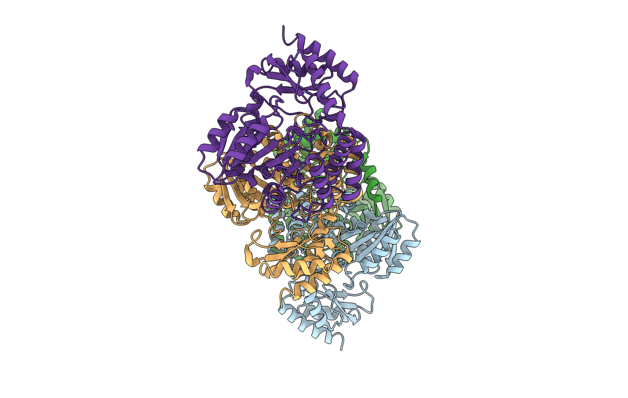
3WY7 image
Deposition Date 2014-08-20
Release Date 2014-12-17
Last Version Date 2023-11-08
Entry Detail
PDB ID:
3WY7
Keywords:
Title:
Crystal structure of Mycobacterium smegmatis 7-Keto-8-aminopelargonic acid (KAPA) synthase BioF
Biological Source:
Source Organism:
Method Details:
Experimental Method:
Resolution:
2.30 Å
R-Value Free:
0.27
R-Value Work:
0.24
R-Value Observed:
0.24
Space Group:
P 1 21 1
Macromolecular Entities
Protein Blast
Polymer Type:polypeptide(L)
Molecule:8-amino-7-oxononanoate synthase
Gene (Uniprot):bioF
Chain IDs:A, B, C, D
Chain Length:404
Number of Molecules:4
Biological Source:Mycobacterium smegmatis str. MC2 155
Primary Citation
Structure and function of Mycobacterium smegmatis 7-keto-8-aminopelargonic acid (KAPA) synthase
Int.J.Biochem.Cell Biol. 58C 71 80 (2014)
PMID: 25462832 DOI: 10.1016/j.biocel.2014.11.006

Abstact

The biotin biosynthesis pathway is an attractive target for development of novel drugs against mycobacterial pathogens, however there are as yet no suitable inhibitors that target this pathway in mycobacteria. 7-Keto-8-aminopelargonic acid synthase (KAPA synthase, BioF) is the enzyme which catalyzes the first committed step of the biotin synthesis pathway, but both its structure and function in mycobacteria remain unresolved. Here we present the crystal structure of Mycobacterium smegmatis BioF (MsBioF). The structure reveals an incomplete dimer, and the active site organization is similar to, but distinct from Escherichia coli 8-amino-7-oxononanoate synthase (EcAONS), the E. coli homologue of BioF. To investigate the influence of structural characteristics on the function of MsBioF, we deleted bioF in M. smegmatis and confirmed that BioF is required for growth in the absence of exogenous biotin. Based on structural and mutagenesis studies, we confirmed that pyridoxal 5'-phosphate (PLP) binding site residues His129, Lys235 and His200 are essential for MsBioF activity in vivo and residue Glu171 plays an important, but not essential role in MsBioF activity. The N-terminus (residues 1-37) is also essential for MsBioF activity in vivo. The structure and function of MsBioF reported here provides further insights for developing new anti-tuberculosis inhibitors aimed at the biotin synthesis pathway.

Legend

Protein

Chemical

Disease

Primary Citation of related structures