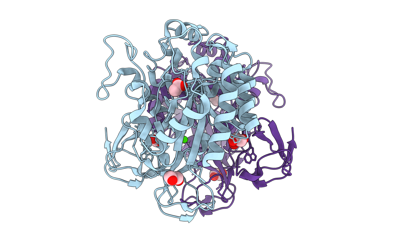
3WX7 image
Deposition Date 2014-07-18
Release Date 2014-12-03
Last Version Date 2024-10-30
Entry Detail
PDB ID:
3WX7
Keywords:
Title:
Crystal structure of COD
Biological Source:
Source Organism:
Method Details:
Experimental Method:
Resolution:
1.35 Å
R-Value Free:
0.19
R-Value Work:
0.17
R-Value Observed:
0.17
Space Group:
P 21 21 21
Macromolecular Entities
Polymer Type:polypeptide(L)
Molecule:Chitin oligosaccharide deacetylase
Gene (Uniprot):cod1
Chain IDs:A, B
Chain Length:402
Number of Molecules:2
Biological Source:Vibrio parahaemolyticus
Primary Citation
Structure-based analysis of domain function of chitin oligosaccharide deacetylase from Vibrio parahaemolyticus.
FEBS Lett. 589 145 151 (2015)
PMID: 25479092 DOI: 10.1016/j.febslet.2014.11.039

Abstact

The X-ray crystal structure of chitin oligosaccharide deacetylase from Vibrio parahaemolyticus (Vp-COD) was determined at an 1.35 Å resolution. The amino acid sequence and structure of Vp-COD show that the enzyme comprises one polysaccharide deacetylase domain (PDD) and two carbohydrate-binding domains (CBDs). On the basis of a chitin-binding assay with Vp-COD and its CBDs-deleted mutant, it was confirmed that CBDs can adhere to chitin. The catalytic activity of the CBDs-deleted mutant was only mildly depressed compared with that of Vp-COD, indicating that CBDs are unlikely to affect the configuration of the active center residues in active site of PDD.

Legend

Protein

Chemical

Disease

Primary Citation of related structures