3WWQ image
Deposition Date 2014-06-23
Release Date 2015-05-13
Last Version Date 2023-11-08
Entry Detail
PDB ID:
3WWQ
Title:
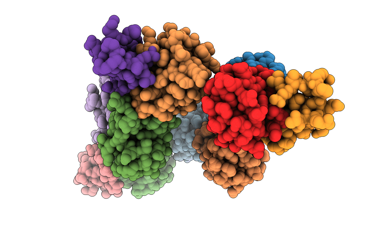
Crystal structure of FAAP20 UBZ domain in complex with Lys63-linked diubiquitin
Biological Source:
Source Organism:
Mus musculus (Taxon ID: 10090)
Homo sapiens (Taxon ID: 9606)
Host Organism:
Method Details:
Experimental Method:
Resolution:
1.90 Å
R-Value Free:
0.23
R-Value Work:
0.20
R-Value Observed:
0.20
Space Group:
P 1 21 1
Macromolecular Entities
Polymer Type:polypeptide(L)
Molecule:Ubiquitin
Gene (Uniprot):Ubc
Mutations:K63R
Chain IDs:A, D, G, J
Chain Length:76
Number of Molecules:4
Biological Source:Mus musculus
Polymer Type:polypeptide(L)
Molecule:Ubiquitin
Gene (Uniprot):Ubc
Chain IDs:B, E, H, K
Chain Length:77
Number of Molecules:4
Biological Source:Mus musculus
Polymer Type:polypeptide(L)
Molecule:Fanconi anemia-associated protein of 20 kDa
Gene (Uniprot):FAAP20
Chain IDs:C, F, I, L
Chain Length:44
Number of Molecules:4
Biological Source:Homo sapiens
Ligand Molecules
Primary Citation
Structural Basis for Ubiquitin Recognition by Ubiquitin-Binding Zinc Finger of FAAP20
Plos One 10 e0120887 e0120887 (2015)
PMID: 25799058 DOI: 10.1371/journal.pone.0120887

Abstact

Several ubiquitin-binding zinc fingers (UBZs) have been reported to preferentially bind K63-linked ubiquitin chains. In particular, the UBZ domain of FAAP20 (FAAP20-UBZ), a member of the Fanconi anemia core complex, seems to recognize K63-linked ubiquitin chains, in order to recruit the complex to DNA interstrand crosslinks and mediate DNA repair. By contrast, it is reported that the attachment of a single ubiquitin to Rev1, a translesion DNA polymerase, increases binding of Rev1 to FAAP20. To clarify the specificity of FAAP20-UBZ, we determined the crystal structure of FAAP20-UBZ in complex with K63-linked diubiquitin at 1.9 Å resolution. In this structure, FAAP20-UBZ interacts only with one of the two ubiquitin moieties. Consistently, binding assays using surface plasmon resonance spectrometry showed that FAAP20-UBZ binds ubiquitin and M1-, K48- and K63-linked diubiquitin chains with similar affinities. Residues in the vicinity of Ala168 within the α-helix and the C-terminal Trp180 interact with the canonical Ile44-centered hydrophobic patch of ubiquitin. Asp164 within the α-helix and the C-terminal loop mediate a hydrogen bond network, which reinforces ubiquitin-binding of FAAP20-UBZ. Mutations of the ubiquitin-interacting residues disrupted binding to ubiquitin in vitro and abolished the accumulation of FAAP20 to DNA damage sites in vivo. Finally, structural comparison among FAAP20-UBZ, WRNIP1-UBZ and RAD18-UBZ revealed distinct modes of ubiquitin binding. UBZ family proteins could be divided into at least three classes, according to their ubiquitin-binding modes.

Legend

Protein

Chemical

Disease

Primary Citation of related structures