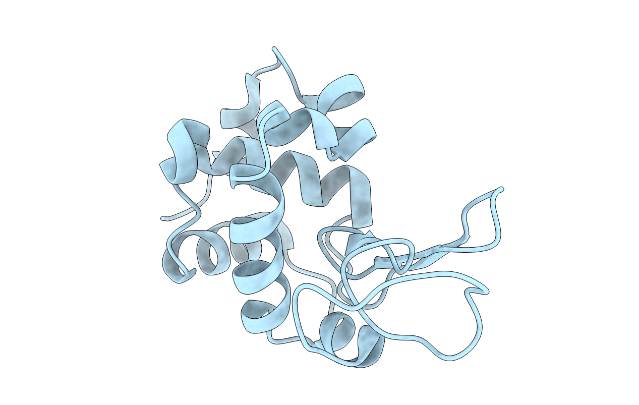
3WW6 image
Deposition Date 2014-06-17
Release Date 2015-06-17
Last Version Date 2024-10-16
Entry Detail
PDB ID:
3WW6
Keywords:
Title:
Crystal Structure of hen egg white lysozyme mutant N46D/D52S
Biological Source:
Source Organism:
Gallus gallus (Taxon ID: 9031)
Host Organism:
Method Details:
Experimental Method:
Resolution:
1.53 Å
R-Value Free:
0.20
R-Value Work:
0.18
R-Value Observed:
0.18
Space Group:
P 43 21 2
Macromolecular Entities
Polymer Type:polypeptide(L)
Molecule:Lysozyme C
Gene (Uniprot):LYZ
Mutations:N46D, D52S
Chain IDs:A
Chain Length:129
Number of Molecules:1
Biological Source:Gallus gallus
Ligand Molecules
Primary Citation
Effect on catalysis by replacement of catalytic residue from hen egg white lysozyme to Venerupis philippinarum lysozyme.
Protein Sci. 25 1637 1647 (2016)
PMID: 27291073 DOI: 10.1002/pro.2966

Abstact

Asn46Asp/Asp52Ser or Asn46Glu/Asp52Ser hen egg white lysozyme (HEL) mutant was designed by introducing the substituted catalytic residue Asp46 or Glu46, respectively, based on Venerupis philippinarum (Vp) lysozyme structure as a representative of invertebrate-type (i-type) lyzozyme. These mutations restored the bell-shaped pH-dependency of the enzyme activity from the sigmoidal pH-dependency observed for the Asp52Ser mutant. Furthermore both lysozyme mutants possessed retaining mechanisms like Vp lysozyme and HEL. The Asn46Glu/Asp52Ser mutant, which has a shorter distance between two catalytic residues, formed a glycosyl adduct in the reaction with the N-acetylglucosamine oligomer. Furthermore, we found the accelerated turnover through its glycosyl adduct formation and decomposition. The turnover rate estimated from the glycosyl formation and decomposition rates was only 20% of the observed hydrolysis rate of the substrate. Based on these results, we discussed the catalytic mechanism of lysozymes.

Legend

Protein

Chemical

Disease

Primary Citation of related structures