Deposition Date
2014-05-22
Release Date
2015-06-24
Last Version Date
2023-11-08
Entry Detail
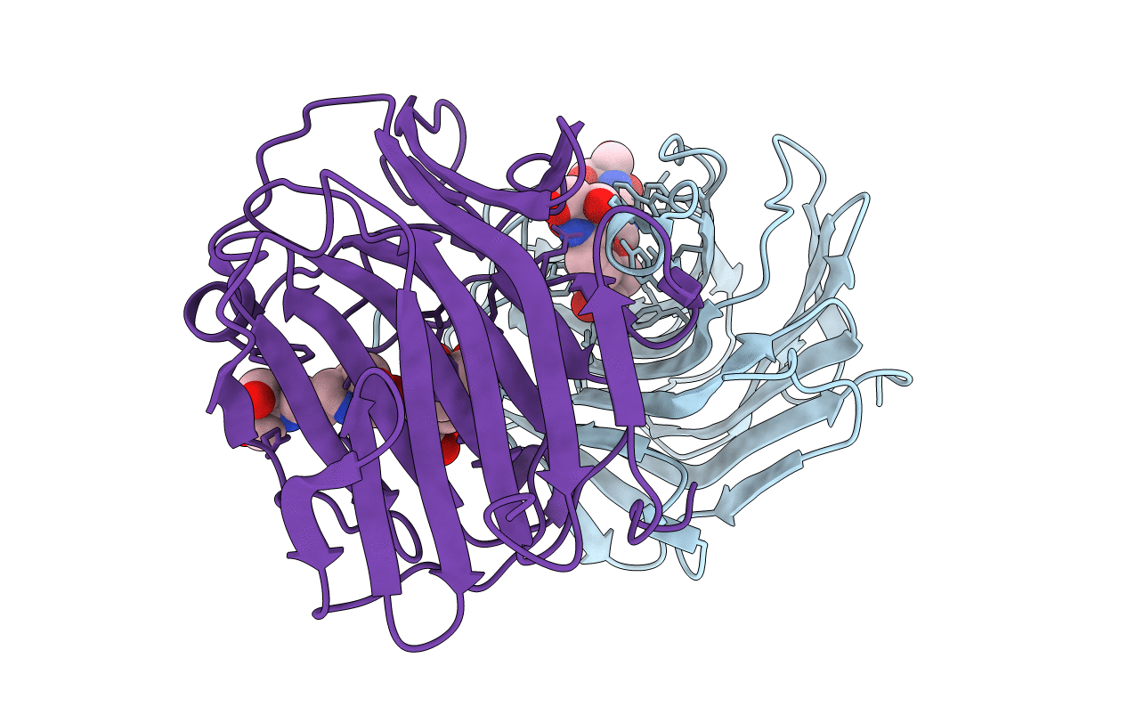
Biological Source:
Source Organism(s):
Clostridium thermocellum (Taxon ID: 203119)
Expression System(s):
Method Details:
Experimental Method:
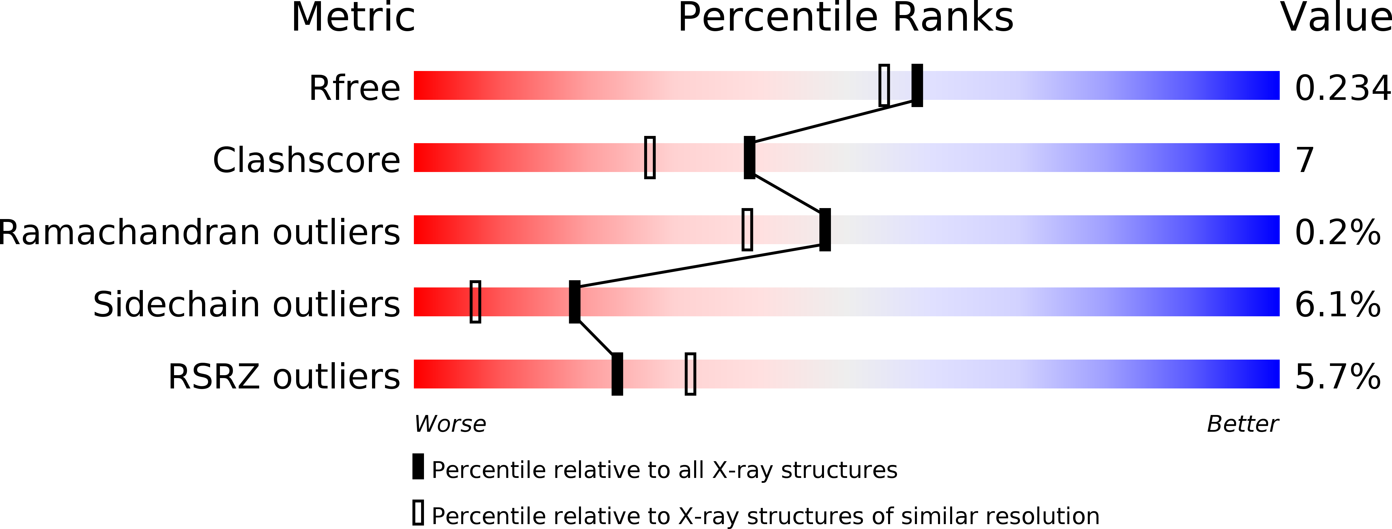
Resolution:
1.95 Å
R-Value Free:
0.23
R-Value Work:
0.18
R-Value Observed:
0.19
Space Group:
P 31 2 1