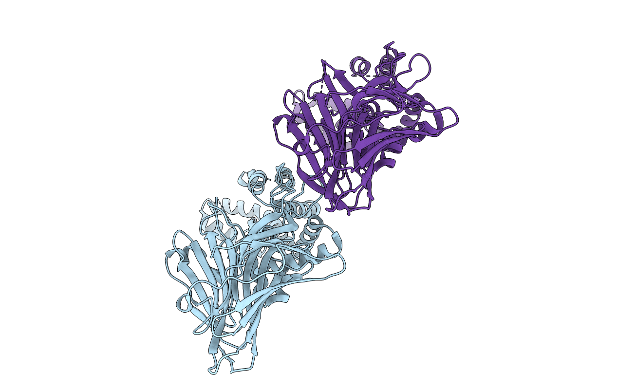
3WVF image
Entry Detail
PDB ID:
3WVF
Keywords:
Title:
Crystal structure of YidC from Escherichia coli
Biological Source:
Source Organism:
Host Organism:
PDB Version:
Deposition Date:
2014-05-20
Release Date:
2014-12-17
Method Details:
Experimental Method:
Resolution:
3.20 Å
R-Value Free:
0.27
R-Value Work:
0.22
R-Value Observed:
0.22
Space Group:
P 1
Macromolecular Entities
Polymer Type:polypeptide(L)
Description:Membrane protein insertase YidC
Chain IDs:A, B
Chain Length:551
Number of Molecules:2
Biological Source:Escherichia coli
Ligand Molecules
Primary Citation
Crystal structure of Escherichia coli YidC, a membrane protein chaperone and insertase
Sci Rep 4 7299 7299 (2014)
PMID: 25466392 DOI: 10.1038/srep07299

Abstact

Bacterial YidC, an evolutionally conserved membrane protein, functions as a membrane protein chaperone in cooperation with the Sec translocon and as an independent insertase for membrane proteins. In Gram-negative bacteria, the transmembrane and periplasmic regions of YidC interact with the Sec proteins, forming a multi-protein complex for Sec-dependent membrane protein integration. Here, we report the crystal structure of full-length Escherichia coli YidC. The structure reveals that a hydrophilic groove, formed by five transmembrane helices, is a conserved structural feature of YidC, as compared to the previous YidC structure from Bacillus halodurans, which lacks a periplasmic domain. Structural mapping of the substrate- or Sec protein-contact sites suggested the importance of the groove for the YidC functions as a chaperone and an insertase, and provided structural insight into the multi-protein complex.

Legend

Protein

Chemical

Disease

Primary Citation of related structures