3WSV image
Deposition Date 2014-03-27
Release Date 2014-09-10
Last Version Date 2023-11-08
Entry Detail
PDB ID:
3WSV
Keywords:
Title:
Crystal structure of minor L-lactate dehydrogenase from Enterococcus mundtii in the ligands-unbound form
Biological Source:
Source Organism:
Host Organism:
Method Details:
Experimental Method:
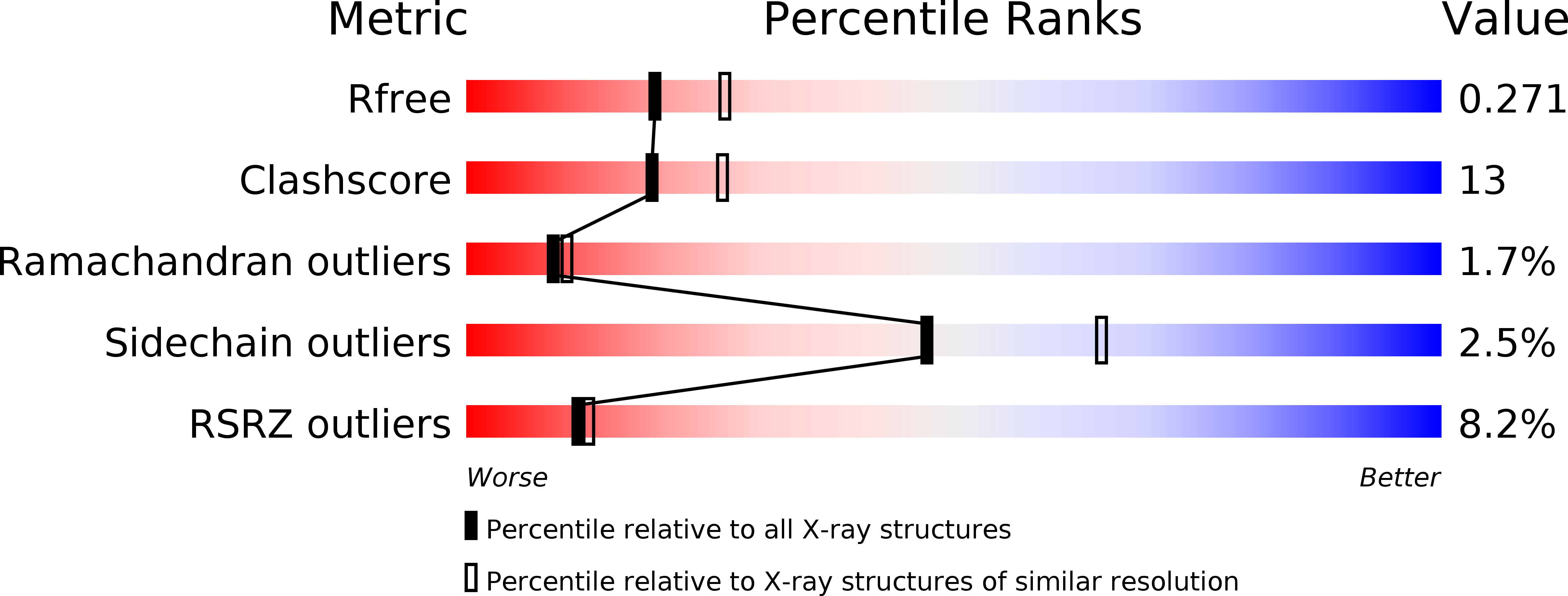
Resolution:
2.38 Å
R-Value Free:
0.27
R-Value Work:
0.23
R-Value Observed:
0.23
Space Group:
P 21 21 21
Macromolecular Entities
Polymer Type:polypeptide(L)
Molecule:L-lactate dehydrogenase
Chain IDs:A, B, C, D
Chain Length:322
Number of Molecules:4
Biological Source:Enterococcus mundtii
Ligand Molecules
Primary Citation
An alternative allosteric regulation mechanism of an acidophilic l-lactate dehydrogenase from Enterococcus mundtii 15-1A.
Febs Open Bio 4 834 847 (2014)
PMID: 25379380 DOI: 10.1016/j.fob.2014.08.006

Abstact

A plant-derived Enterococcus mundtii 15-1A, that has been previously isolated from Brassica rapa L. subsp. nipposinica (L.H. Bailey) Hanelt var. linearifolia by our group, possesses two kinds of l-lactate dehydrogenase (l-LDH): LDH-1 and LDH-2. LDH-1 was activated under low concentration of fluctose-1,6-bisphosphate (FBP) at both pH 5.5 and 7.5. Although LDH-2 was also activated under the low concentration of FBP at pH 5.5, a high concentration of FBP is necessary to activate it at pH 7.5. The present study shows the crystal structures of the acidophilic LDH-2 in a complex with and without FBP and NADH. Although the tertiary structure of the ligands-bound LDH-2 is similar to that of the active form of other bacterial l-LDHs, the structure without the ligands is different from that of any other previously determined l-LDHs. Major structural alterations between the two structures of LDH-2 were observed at two regions in one subunit. At the N-terminal parts of the two regions, the ligands-bound form takes an α-helical structure, while the form without ligands displays more disordered and extended structures. A vacuum-ultraviolet circular dichroism analysis showed that the α-helix content of LDH-2 in solution is approximately 30% at pH 7.5, which is close to that in the crystal structure of the form without ligands. A D241N mutant of LDH-2, which was created by us to easily form an α-helix at one of the two parts, exhibited catalytic activity even in the absence of FBP at both pH 5.5 and 7.5.

Legend

Protein

Chemical

Disease

Primary Citation of related structures
Feedback Form
Name
Email
Institute
Feedback