
Deposition Date
2014-03-26
Release Date
2015-05-20
Last Version Date
2023-11-08
Entry Detail
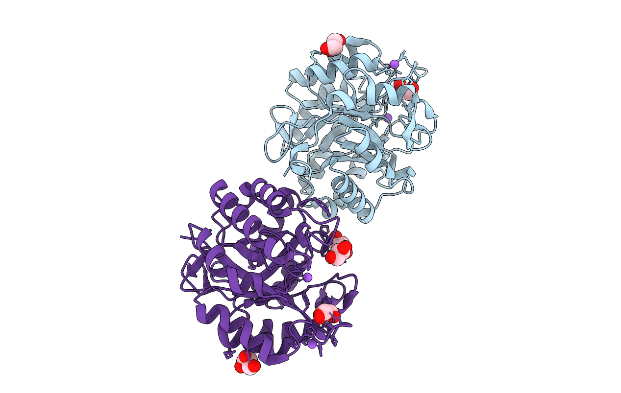
PDB ID:
3WSU
Keywords:
Title:
Crystal structure of beta-mannanase from Streptomyces thermolilacinus
Biological Source:
Source Organism:
Streptomyces thermolilacinus (Taxon ID: 285540)
Host Organism:
Method Details:
Experimental Method:
Resolution:
1.60 Å
R-Value Free:
0.19
R-Value Work:
0.16
R-Value Observed:
0.16
Space Group:
P 21 21 21
