3WSR image
Deposition Date 2014-03-20
Release Date 2014-10-22
Last Version Date 2024-11-20
Entry Detail
PDB ID:
3WSR
Title:
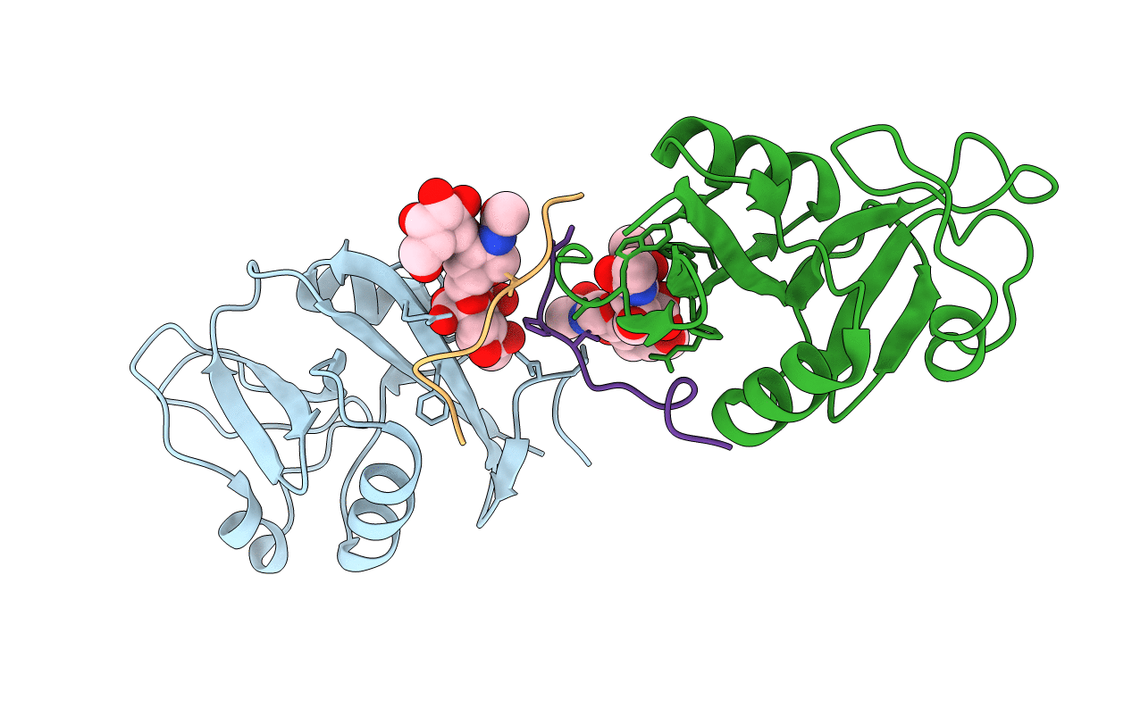
Crystal structure of CLEC-2 in complex with O-glycosylated podoplanin
Biological Source:
Source Organism:
Homo sapiens (Taxon ID: 9606)
Host Organism:
Method Details:
Experimental Method:
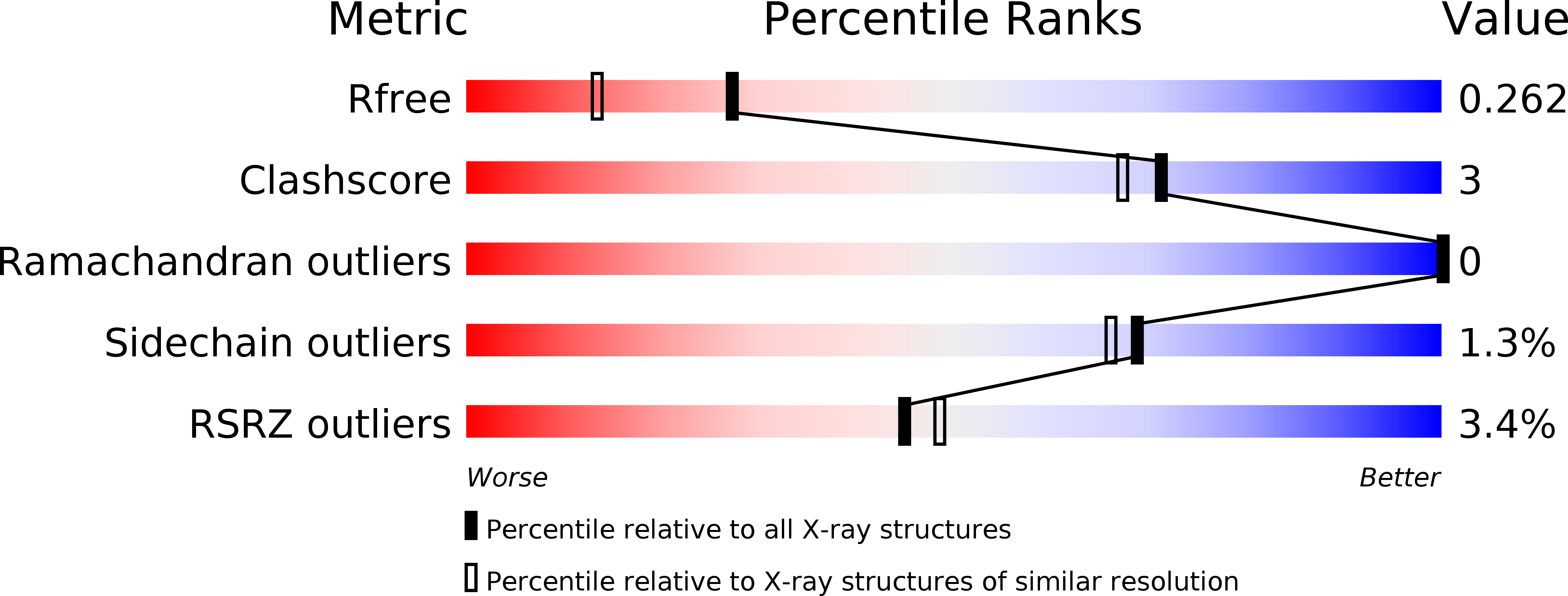
Resolution:
1.91 Å
R-Value Free:
0.25
R-Value Work:
0.21
R-Value Observed:
0.21
Space Group:
P 1 21 1
Macromolecular Entities
Structures with similar UniProt ID
Protein Blast
Polymer Type:polypeptide(L)
Molecule:C-type lectin domain family 1 member B
Gene (Uniprot):CLEC1B
Mutagens:C99S
Chain IDs:A, B
Chain Length:128
Number of Molecules:2
Biological Source:Homo sapiens
Structures with similar UniProt ID
Protein Blast
Polymer Type:polypeptide(L)
Molecule:Peptide from Podoplanin
Gene (Uniprot):PDPN
Chain IDs:C, D
Chain Length:18
Number of Molecules:2
Biological Source:Homo sapiens
Modified Residue
Compound ID Chain ID Parent Comp ID Details 2D Image
THR C THR GLYCOSYLATION SITE
Primary Citation
A Platform of C-type Lectin-like Receptor CLEC-2 for Binding O-Glycosylated Podoplanin and Nonglycosylated Rhodocytin
Structure 22 1711 1721 (2014)
PMID: 25458834 DOI: 10.1016/j.str.2014.09.009

Abstact

Podoplanin is a transmembrane O-glycoprotein that binds to C-type lectin-like receptor 2 (CLEC-2). The O-glycan-dependent interaction seems to play crucial roles in various biological processes, such as platelet aggregation. Rhodocytin, a snake venom, also binds to CLEC-2 and aggregates platelets in a glycan-independent manner. To elucidate the structural basis of the glycan-dependent and independent interactions, we performed comparative crystallographic studies of podoplanin and rhodocytin in complex with CLEC-2. Both podoplanin and rhodocytin bind to the noncanonical "side" face of CLEC-2. There is a common interaction mode between consecutive acidic residues on the ligands and the same arginine residues on CLEC-2. Other interactions are ligand-specific. Carboxyl groups from the sialic acid residue on podoplanin and from the C terminus of the rhodocytin α subunit interact differently at this "second" binding site on CLEC-2. The unique and versatile binding modes open a way to understand the functional consequences of CLEC-2-ligand interactions.

Legend

Protein

Chemical

Disease

Primary Citation of related structures
Feedback Form
Name
Email
Institute
Feedback