3WOA image
Deposition Date 2013-12-25
Release Date 2015-04-29
Last Version Date 2023-11-08
Entry Detail
PDB ID:
3WOA
Title:
Crystal structure of lambda repressor (1-45) fused with maltose-binding protein
Biological Source:
Source Organism:
Host Organism:
Method Details:
Experimental Method:
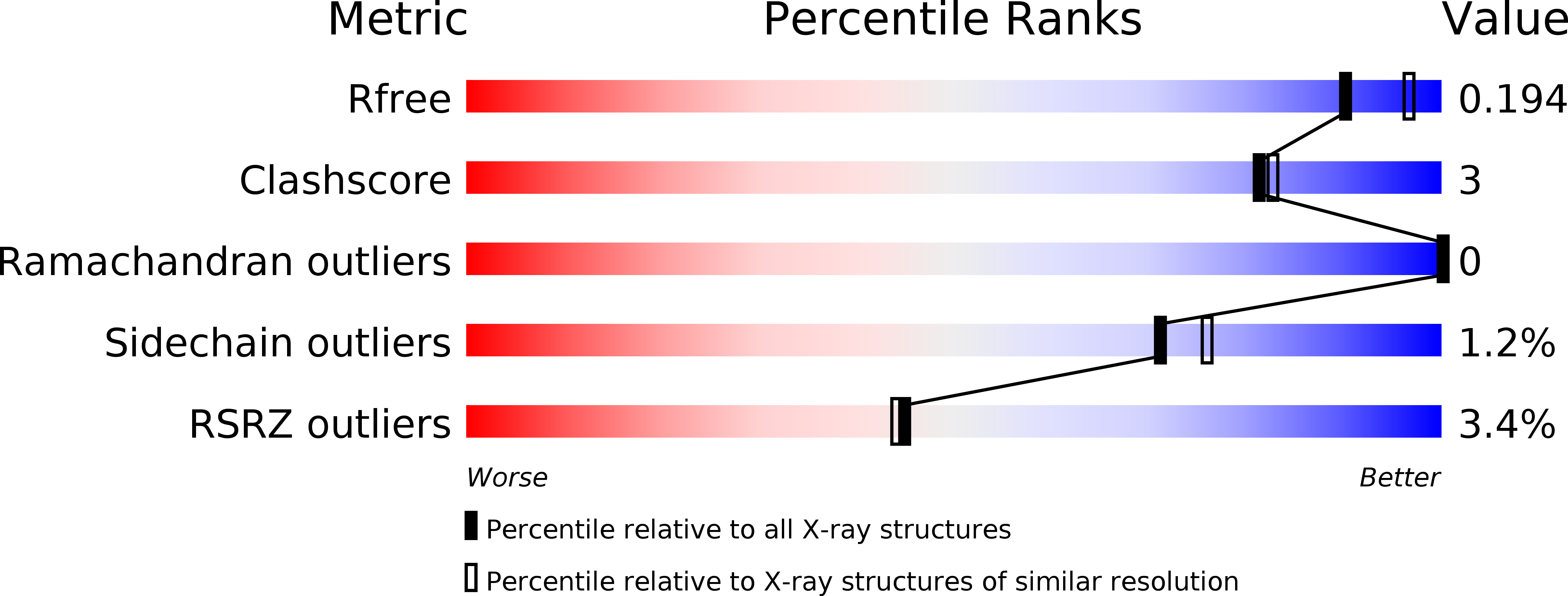
Resolution:
2.00 Å
R-Value Free:
0.19
R-Value Work:
0.15
R-Value Observed:
0.15
Space Group:
P 21 21 2
Macromolecular Entities
Protein Blast
Polymer Type:polypeptide(L)
Molecule:Repressor protein CI, Maltose-binding periplasmic protein
Gene (Uniprot):cI, malE
Mutagens:N416R
Chain IDs:A
Chain Length:417
Number of Molecules:1
Biological Source:Enterobacteria phage lambda, Escherichia coli
Peptide-like Molecules
PRD_900001
Primary Citation
Co-translational folding of alpha-helical proteins: structural studies of intermediate-length variants of the lambda repressor.
FEBS Open Bio 8 1312 1321 (2018)
PMID: 30087834 DOI: 10.1002/2211-5463.12480

Abstact

UNLABELLED Nascent polypeptide chains fold cotranslationally, but the atomic-level details of this process remain unknown. Here, we report crystallographic, de novo modeling, and spectroscopic studies of intermediate-length variants of the λ repressor N-terminal domain. Although the ranges of helical regions of the half-length variant were almost identical to those of the full-length protein, the relative orientations of these helices in the intermediate-length variants differed. Our results suggest that cotranslational folding of the λ repressor initially forms a helical structure with a transient conformation, as in the case of a molten globule state. This conformation subsequently matures during the course of protein synthesis. DATABASE Structural data are available in the PDB under the accession numbers http://www.rcsb.org/pdb/search/structidSearch.do?structureId=5ZCA and http://www.rcsb.org/pdb/search/structidSearch.do?structureId=3WOA.

Legend

Protein

Chemical

Disease

Primary Citation of related structures