3WNU image
Deposition Date 2013-12-17
Release Date 2014-03-12
Last Version Date 2024-03-20
Entry Detail
PDB ID:
3WNU
Keywords:
Title:
The crystal structure of catalase-peroxidase, KatG, from Synechococcus PCC7942
Biological Source:
Source Organism:
Host Organism:
Method Details:
Experimental Method:
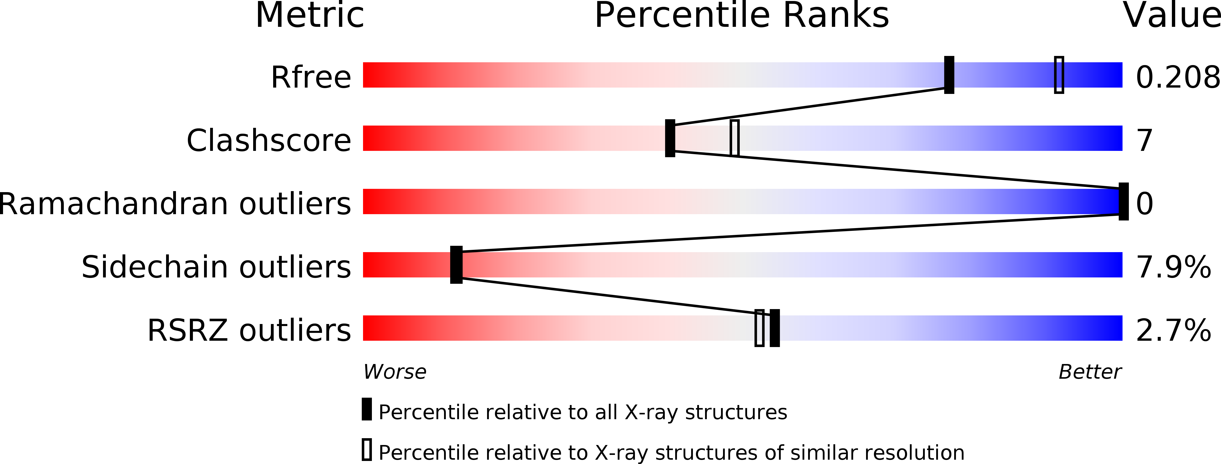
Resolution:
2.20 Å
R-Value Free:
0.20
R-Value Work:
0.16
R-Value Observed:
0.16
Space Group:
P 41 21 2
Macromolecular Entities
Structures with similar UniProt ID
Protein Blast
Polymer Type:polypeptide(L)
Molecule:Catalase-peroxidase
Gene (Uniprot):katG
Chain IDs:A
Chain Length:720
Number of Molecules:1
Biological Source:Synechococcus elongatus
Primary Citation
The 2.2 angstrom resolution structure of the catalase-peroxidase KatG from Synechococcus elongatus PCC7942.
Acta Crystallogr.,Sect.F 70 288 293 (2014)
PMID: 24598912 DOI: 10.1107/S2053230X14002052

Abstact

The crystal structure of catalase-peroxidase from Synechococcus elongatus PCC7942 (SeKatG) was solved by molecular replacement and refined to an Rwork of 16.8% and an Rfree of 20.6% at 2.2 Å resolution. The asymmetric unit consisted of only one subunit of the catalase-peroxidase molecule, including a protoporphyrin IX haem moiety and two sodium ions. A typical KatG covalent adduct was formed, Met248-Tyr222-Trp94, which is a key structural element for catalase activity. The crystallographic equivalent subunit was created by a twofold symmetry operation to form the functional dimer. The overall structure of the dimer was quite similar to other KatGs. One sodium ion was located close to the proximal Trp314. The location and configuration of the proximal cation site were very similar to those of typical peroxidases such as ascorbate peroxidase. These features may provide a structural basis for the behaviour of the radical localization/delocalization during the course of the enzymatic reaction.

Legend

Protein

Chemical

Disease

Primary Citation of related structures