3WMU image
Deposition Date 2013-11-27
Release Date 2014-12-03
Last Version Date 2024-03-20
Entry Detail
PDB ID:
3WMU
Title:
The structure of an anti-cancer lectin mytilec apo-form from the mussel Mytilus galloprovincialis
Biological Source:
Source Organism:
Host Organism:
Method Details:
Experimental Method:
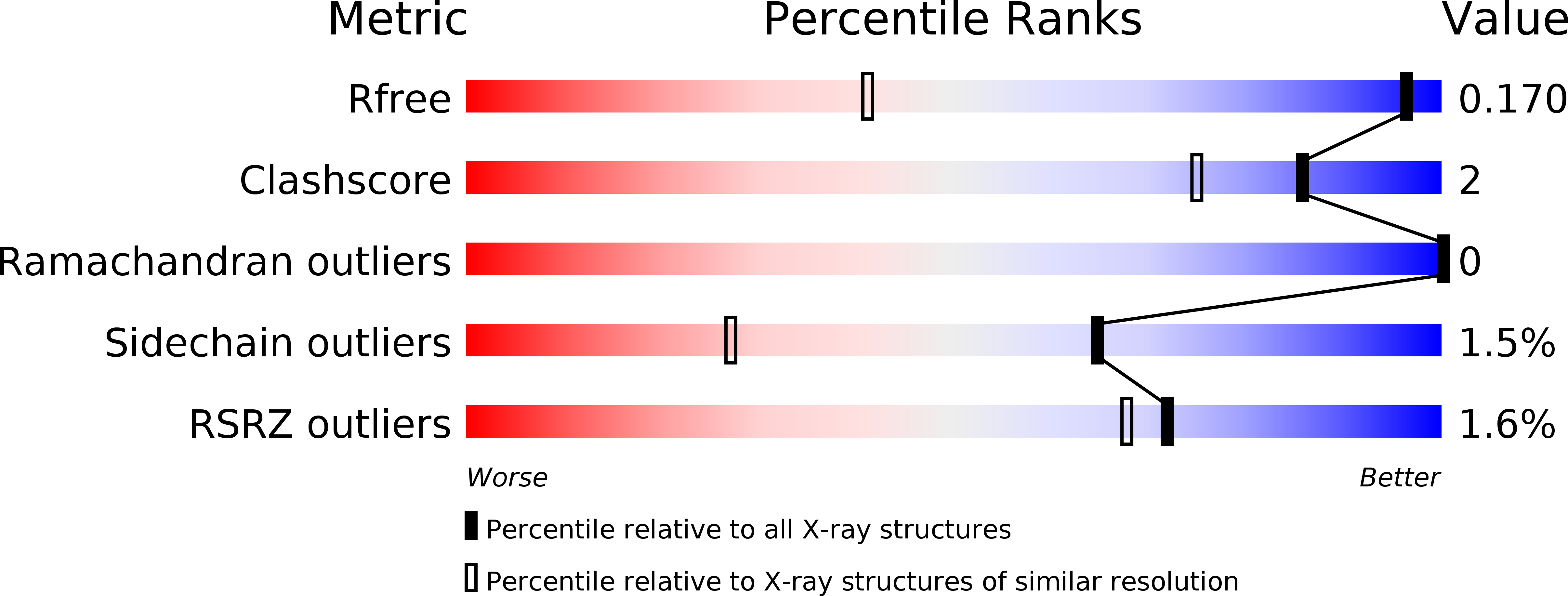
Resolution:
1.10 Å
R-Value Free:
0.16
R-Value Work:
0.13
R-Value Observed:
0.13
Space Group:
P 1 21 1
Macromolecular Entities
Polymer Type:polypeptide(L)
Molecule:Lectin
Mutations:T1A
Chain IDs:A, B
Chain Length:153
Number of Molecules:2
Biological Source:Mytilus galloprovincialis
Ligand Molecules
Primary Citation
Crystal structure of MytiLec, a galactose-binding lectin from the mussel Mytilus galloprovincialis with cytotoxicity against certain cancer cell types
Sci Rep 6 28344 28344 (2016)
PMID: 27321048 DOI: 10.1038/srep28344

Abstact

MytiLec is a lectin, isolated from bivalves, with cytotoxic activity against cancer cell lines that express globotriaosyl ceramide, Galα(1,4)Galβ(1,4)Glcα1-Cer, on the cell surface. Functional analysis shows that the protein binds to the disaccharide melibiose, Galα(1,6)Glc, and the trisaccharide globotriose, Galα(1,4)Galβ(1,4)Glc. Recombinant MytiLec expressed in bacteria showed the same haemagglutinating and cytotoxic activity against Burkitt's lymphoma (Raji) cells as the native form. The crystal structure has been determined to atomic resolution, in the presence and absence of ligands, showing the protein to be a member of the β-trefoil family, but with a mode of ligand binding unique to a small group of related trefoil lectins. Each of the three pseudo-equivalent binding sites within the monomer shows ligand binding, and the protein forms a tight dimer in solution. An engineered monomer mutant lost all cytotoxic activity against Raji cells, but retained some haemagglutination activity, showing that the quaternary structure of the protein is important for its cellular effects.

Legend

Protein

Chemical

Disease

Primary Citation of related structures