3WMT image
Deposition Date 2013-11-25
Release Date 2014-08-06
Last Version Date 2024-10-09
Entry Detail
PDB ID:
3WMT
Keywords:
Title:
Crystal structure of feruloyl esterase B from Aspergillus oryzae
Biological Source:
Source Organism:
Host Organism:
Method Details:
Experimental Method:
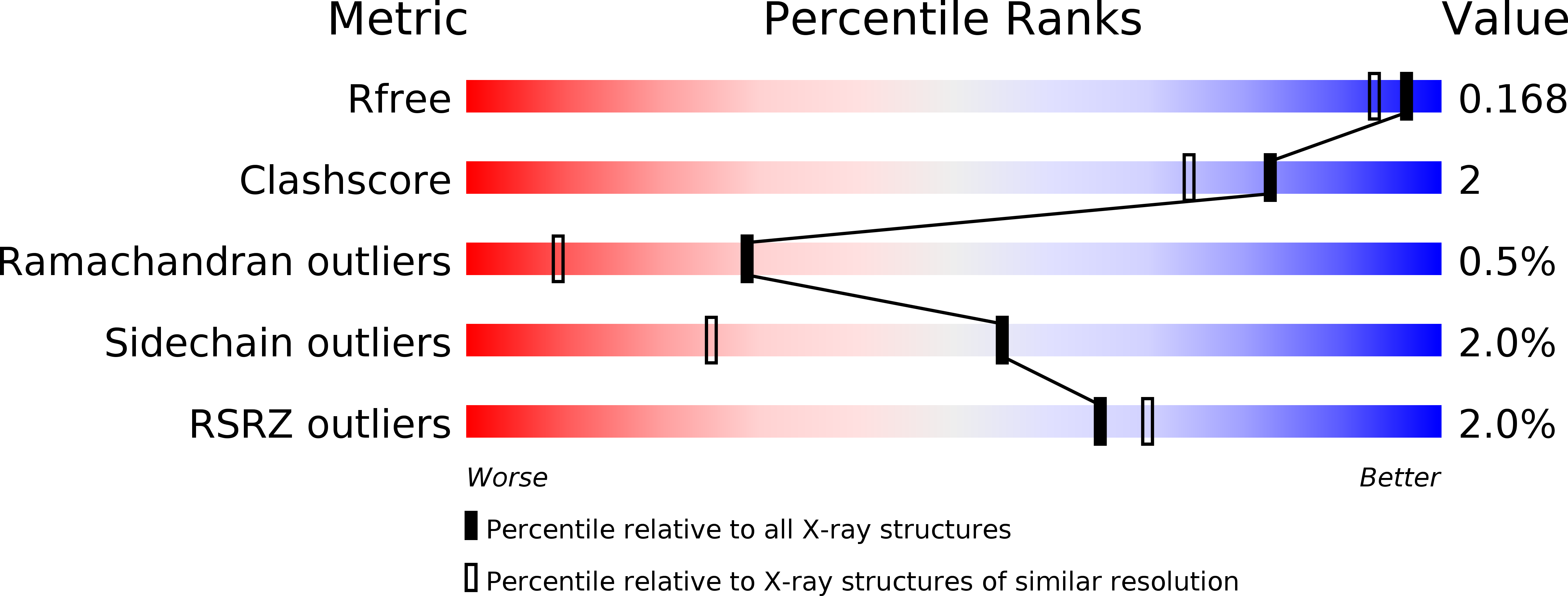
Resolution:
1.50 Å
R-Value Free:
0.16
R-Value Work:
0.14
R-Value Observed:
0.14
Space Group:
P 21 21 2
Macromolecular Entities
Polymer Type:polypeptide(L)
Molecule:Probable feruloyl esterase B-1
Gene (Uniprot):faeB-1
Chain IDs:A, B
Chain Length:522
Number of Molecules:2
Biological Source:Aspergillus oryzae
Modified Residue
Compound ID Chain ID Parent Comp ID Details 2D Image
ASN A ASN GLYCOSYLATION SITE
Primary Citation
Crystal structure of a feruloyl esterase belonging to the tannase family: a disulfide bond near a catalytic triad.
Proteins 82 2857 2867 (2014)
PMID: 25066066 DOI: 10.1002/prot.24649

Abstact

Feruloyl esterase (FAE) catalyzes the hydrolysis of the ferulic and diferulic acids present in plant cell wall polysaccharides, and tannase catalyzes the hydrolysis of tannins to release gallic acid. The fungal tannase family in the ESTHER database contains various enzymes, including FAEs and tannases. Despite the importance of FAEs and tannases in bioindustrial applications, three-dimensional structures of the fungal tannase family members have been unknown. Here, we determined the crystal structure of FAE B from Aspergillus oryzae (AoFaeB), which belongs to the fungal tannase family, at 1.5 Å resolution. AoFaeB consists of a catalytic α/β-hydrolase fold domain and a large lid domain, and the latter has a novel fold. To estimate probable binding models of substrates in AoFaeB, an automated docking analysis was performed. In the active site pocket of AoFaeB, residues responsible for the substrate specificity of the FAE activity were identified. The catalytic triad of AoFaeB comprises Ser203, Asp417, and His457, and the serine and histidine residues are directly connected by a disulfide bond of the neighboring cysteine residues, Cys202 and Cys458. This structural feature, the "CS-D-HC motif," is unprecedented in serine hydrolases. A mutational analysis indicated that the novel structural motif plays essential roles in the function of the active site.

Legend

Protein

Chemical

Disease

Primary Citation of related structures