3WL6 image
Deposition Date 2013-11-08
Release Date 2014-09-24
Last Version Date 2024-10-09
Entry Detail
PDB ID:
3WL6
Keywords:
Title:
Crystal Structure of pOPH Native
Biological Source:
Source Organism:
Pseudomonas sp. (Taxon ID: 306)
Host Organism:
Method Details:
Experimental Method:
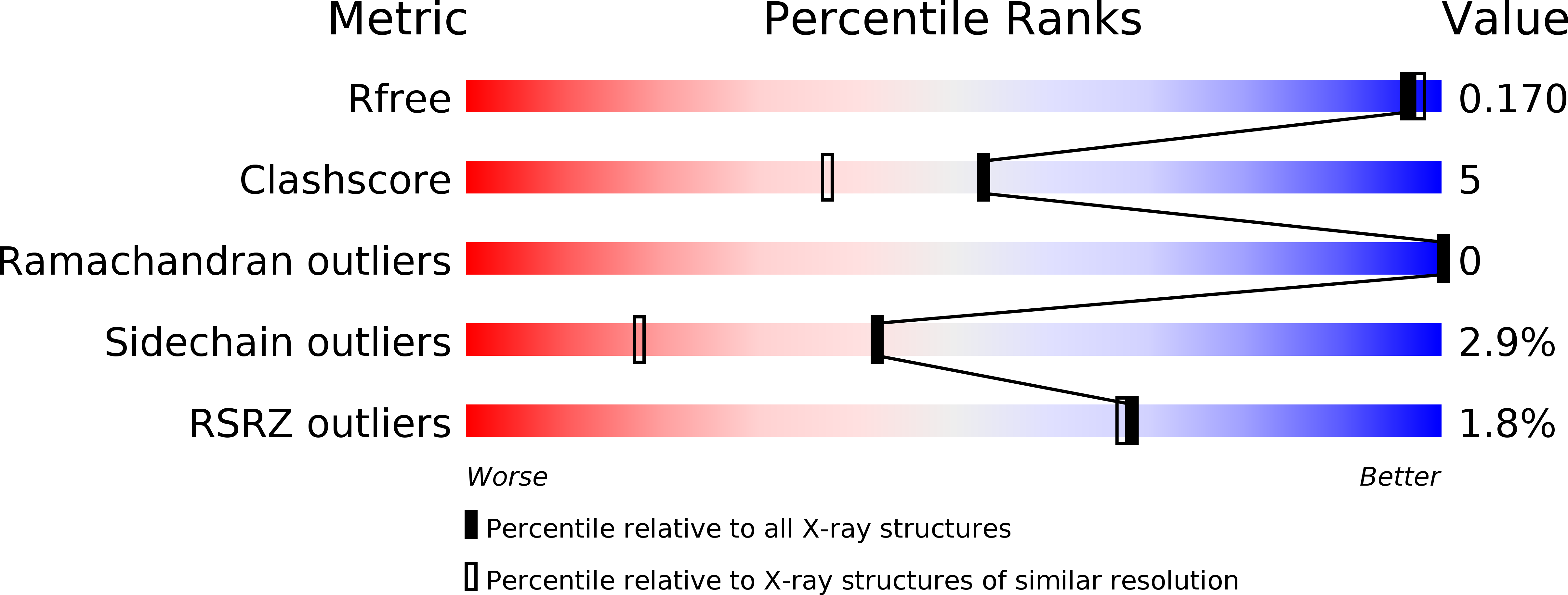
Resolution:
1.60 Å
R-Value Free:
0.17
R-Value Work:
0.15
R-Value Observed:
0.15
Space Group:
P 1
Macromolecular Entities
Structures with similar UniProt ID
Protein Blast
Polymer Type:polypeptide(L)
Molecule:Oxidized polyvinyl alcohol hydrolase
Gene (Uniprot):pvaB
Chain IDs:A, B
Chain Length:364
Number of Molecules:2
Biological Source:Pseudomonas sp.
Ligand Molecules
Primary Citation
Structural insights into enzymatic degradation of oxidized polyvinyl alcohol
Chembiochem 15 1882 1886 (2014)
PMID: 25044912 DOI: 10.1002/cbic.201402166

Abstact

The ever-increasing production and use of polyvinyl alcohol (PVA) threaten our environment. Yet PVA can be assimilated by microbes in two steps: oxidation and cleavage. Here we report novel α/β-hydrolase structures of oxidized PVA hydrolase (OPH) from two known PVA-degrading organisms, Sphingopyxis sp. 113P3 and Pseudomonas sp. VM15C, including complexes with substrate analogues, acetylacetone and caprylate. The active site is covered by a lid-like β-ribbon. Unlike other esterase and amidase, OPH is unique in cleaving the CC bond of β-diketone, although it has a catalytic triad similar to that of most α/β-hydrolases. Analysis of the crystal structures suggests a double-oxyanion-hole mechanism, previously only found in thiolase cleaving β-ketoacyl-CoA. Three mutations in the lid region showed enhanced activity, with potential in industrial applications.

Legend

Protein

Chemical

Disease

Primary Citation of related structures
Feedback Form
Name
Email
Institute
Feedback