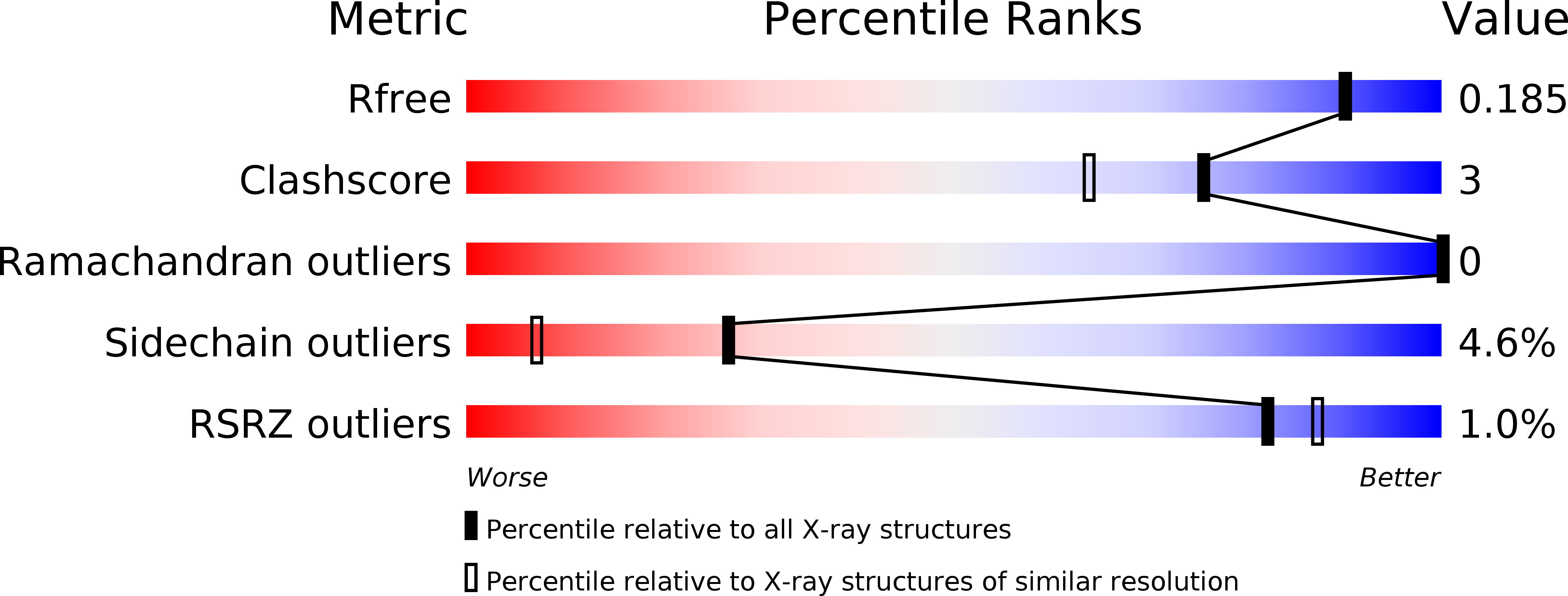
3WKF image
Deposition Date 2013-10-21
Release Date 2013-12-25
Last Version Date 2023-11-08
Entry Detail
PDB ID:
3WKF
Keywords:
Title:
Crystal structure of cellobiose 2-epimerase
Biological Source:
Source Organism:
Host Organism:
Method Details:
Experimental Method:
Resolution:
1.74 Å
R-Value Free:
0.18
R-Value Work:
0.15
R-Value Observed:
0.15
Space Group:
P 21 21 21
Macromolecular Entities
Polymer Type:polypeptide(L)
Molecule:Cellobiose 2-epimerase
Gene (Uniprot):ce
Chain IDs:A
Chain Length:412
Number of Molecules:1
Biological Source:Rhodothermus marinus
Primary Citation
Structural Insights into the Epimerization of beta-1,4-Linked Oligosaccharides Catalyzed by Cellobiose 2-Epimerase, the Sole Enzyme Epimerizing Non-anomeric Hydroxyl Groups of Unmodified Sugars
J.Biol.Chem. 289 3405 3415 (2014)
PMID: 24362032 DOI: 10.1074/jbc.M113.531251

Abstact

Cellobiose 2-epimerase (CE) reversibly converts d-glucose residues into d-mannose residues at the reducing end of unmodified β1,4-linked oligosaccharides, including β-1,4-mannobiose, cellobiose, and lactose. CE is responsible for conversion of β1,4-mannobiose to 4-O-β-d-mannosyl-d-glucose in mannan metabolism. However, the detailed catalytic mechanism of CE is unclear due to the lack of structural data in complex with ligands. We determined the crystal structures of halothermophile Rhodothermus marinus CE (RmCE) in complex with substrates/products or intermediate analogs, and its apo form. The structures in complex with the substrates/products indicated that the residues in the β5-β6 loop as well as those in the inner six helices form the catalytic site. Trp-322 and Trp-385 interact with reducing and non-reducing end parts of these ligands, respectively, by stacking interactions. The architecture of the catalytic site also provided insights into the mechanism of reversible epimerization. His-259 abstracts the H2 proton of the d-mannose residue at the reducing end, and consistently forms the cis-enediol intermediate by facilitated depolarization of the 2-OH group mediated by hydrogen bonding interaction with His-200. His-390 subsequently donates the proton to the C2 atom of the intermediate to form a d-glucose residue. The reverse reaction is mediated by these three histidines with the inverse roles of acid/base catalysts. The conformation of cellobiitol demonstrated that the deprotonation/reprotonation step is coupled with rotation of the C2-C3 bond of the open form of the ligand. Moreover, it is postulated that His-390 is closely related to ring opening/closure by transferring a proton between the O5 and O1 atoms of the ligand.

Legend

Protein

Chemical

Disease

Primary Citation of related structures