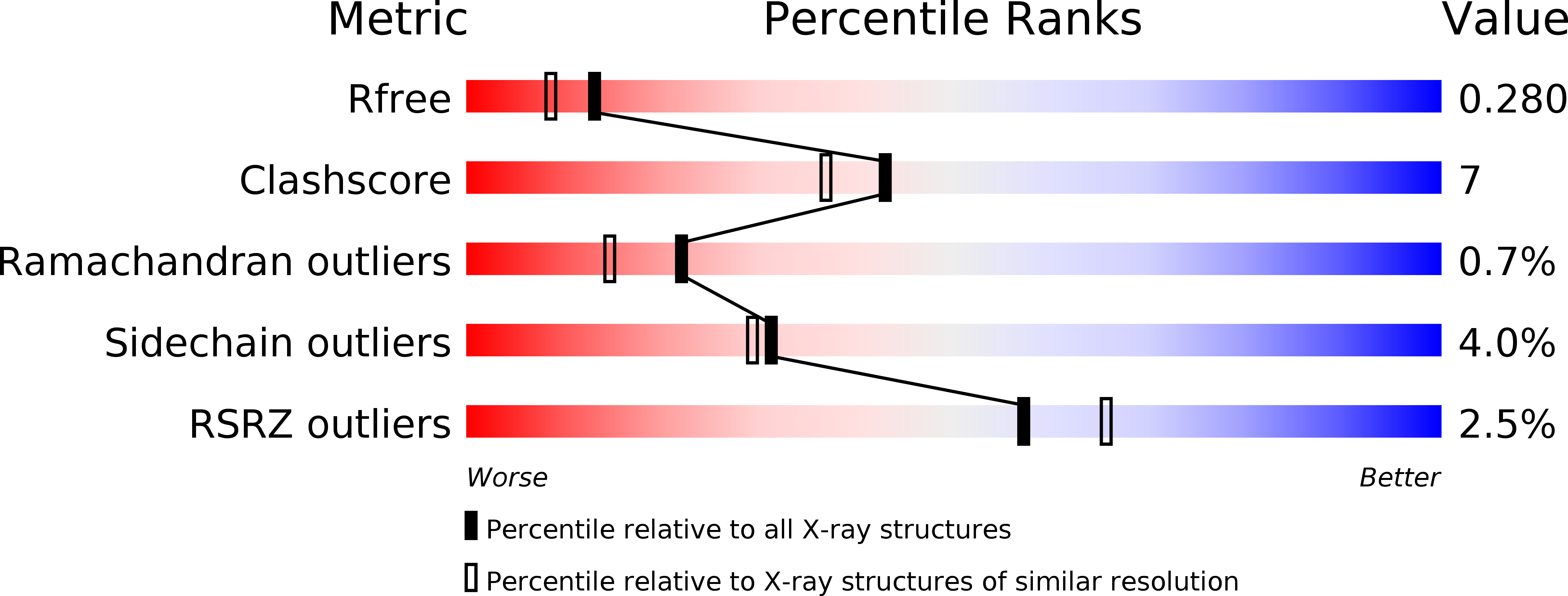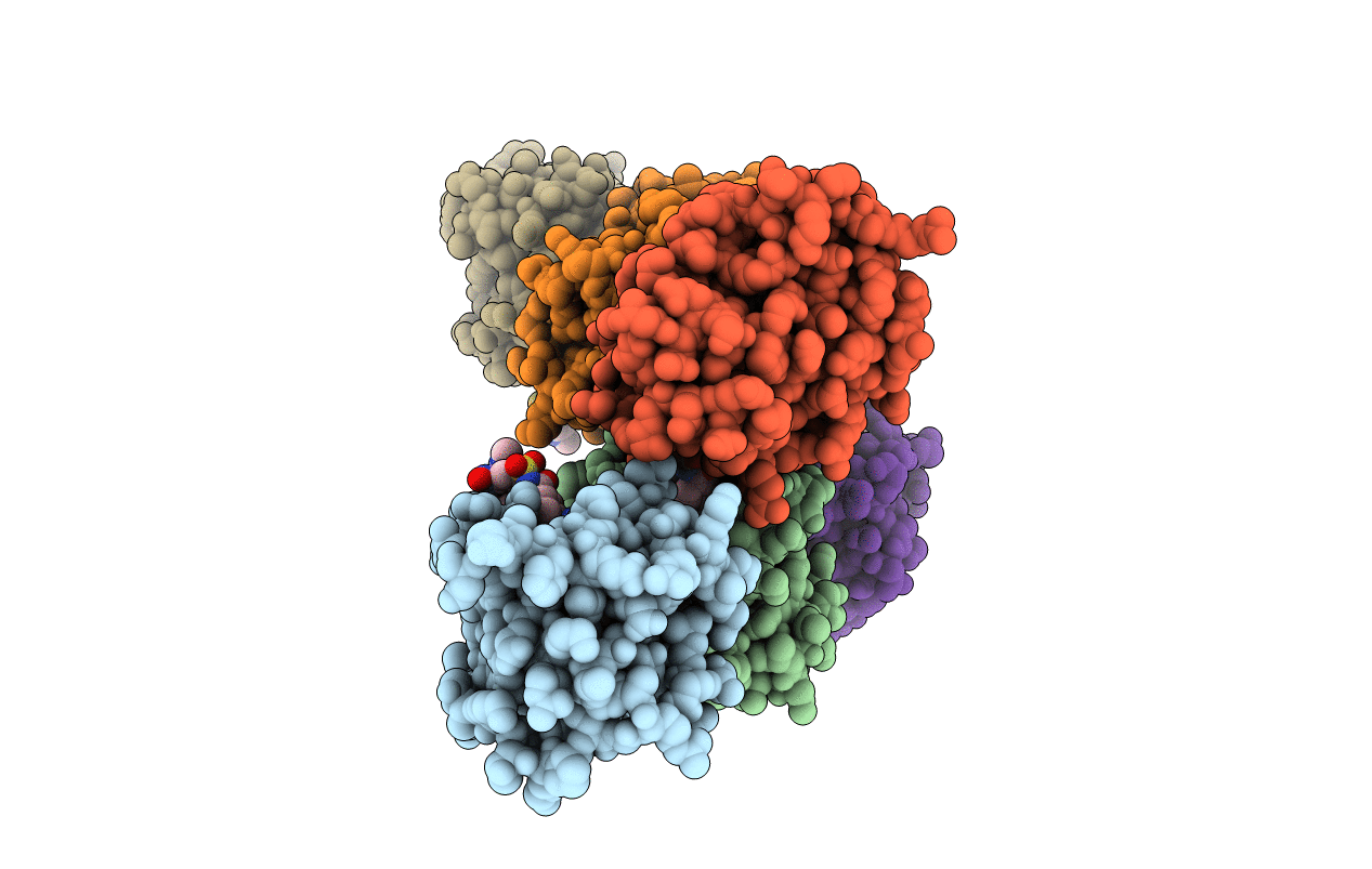
3WIY image
Entry Detail
PDB ID:
3WIY
Keywords:
Title:
Crystal structure of Mcl-1 in complex with compound 10
Biological Source:
Source Organism:
Host Organism:
PDB Version:
Deposition Date:
2013-09-26
Release Date:
2013-11-27
Method Details:
Experimental Method:
Resolution:
2.15 Å
R-Value Free:
0.28
R-Value Work:
0.21
R-Value Observed:
0.21
Space Group:
P 1
Macromolecular Entities
Polymer Type:polypeptide(L)
Description:Induced myeloid leukemia cell differentiation protein Mcl-1
Chain IDs:A, B, C, D, E, F
Chain Length:161
Number of Molecules:6
Biological Source:Homo sapiens
Ligand Molecules
Primary Citation
Discovery of potent Mcl-1/Bcl-xL dual inhibitors by using a hybridization strategy based on structural analysis of target proteins.
J.Med.Chem. 56 9635 9645 (2013)
PMID: 24215352 DOI: 10.1021/jm401170c

Abstact

Mcl-1 and Bcl-xL are crucial regulators of apoptosis, therefore dual inhibitors of both proteins could serve as promising new anticancer drugs. To design Mcl-1/Bcl-xL dual inhibitors, we performed structure-guided analyses of the corresponding selective Mcl-1 and Bcl-xL inhibitors. A cocrystal structure of a pyrazolo[1,5-a]pyridine derivative with Mcl-1 protein was successfully determined and revealed the protein-ligand binding mode. The key structure for Bcl-xL inhibition was further confirmed through the substructural analysis of ABT-263, a representative Bcl-xL/Bcl-2/Bcl-w inhibitor developed by Abbott Laboratories. On the basis of the structural data from this analysis, we designed hybrid compounds by tethering the Mcl-1 and Bcl-xL inhibitors together. The results of X-ray crystallographic analysis of hybrid compound 10 in complexes with both Mcl-1 and Bcl-xL demonstrated its binding mode with each protein. Following further optimization, compound 11 showed potent Mcl-1/Bcl-xL dual inhibitory activity (Mcl-1, IC50 = 0.088 μM; and Bcl-xL, IC50 = 0.0037 μM).

Legend

Protein

Chemical

Disease

Primary Citation of related structures