3WHQ image
Entry Detail
PDB ID:
3WHQ
Title:
Crystal structure of gamma-glutamyltranspeptidase from Bacillus subtilis (crystal soaked for 0 min. in acivicin soln. )
Biological Source:
Source Organism:
Host Organism:
PDB Version:
Deposition Date:
2013-08-30
Release Date:
2014-02-19
Method Details:
Experimental Method:
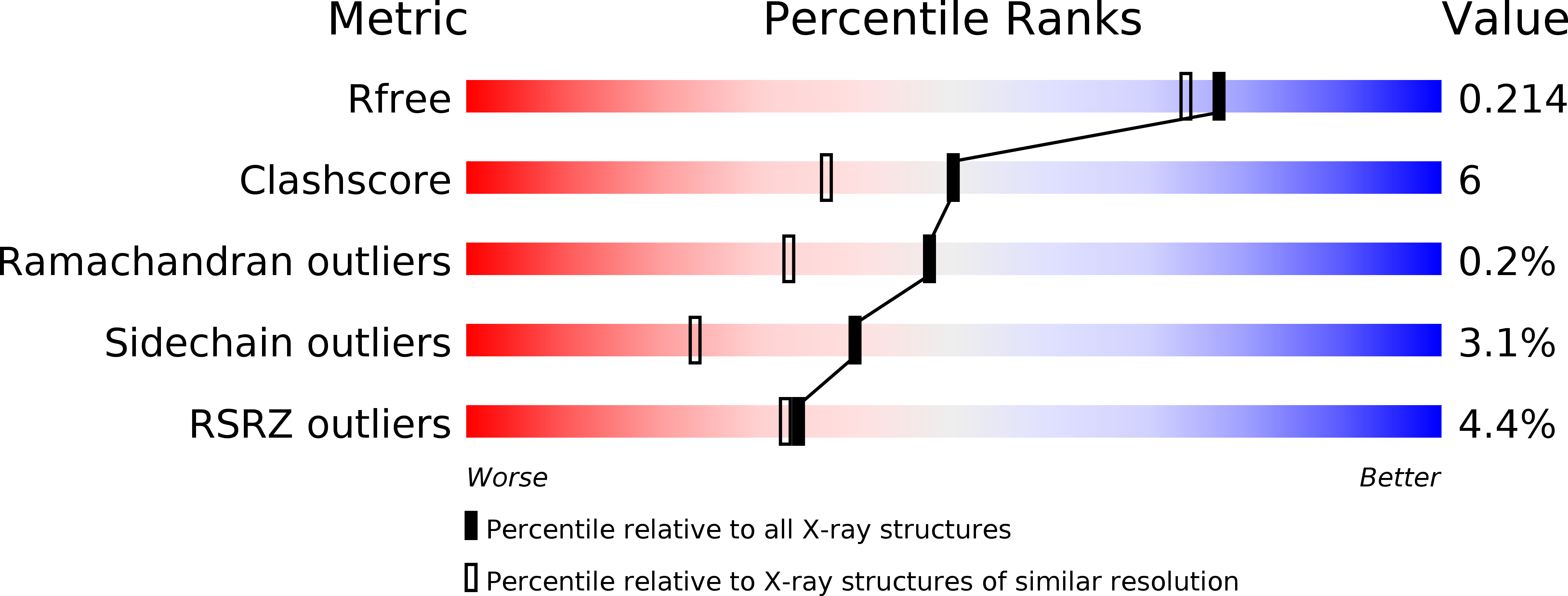
Resolution:
1.85 Å
R-Value Free:
0.21
R-Value Work:
0.17
R-Value Observed:
0.17
Space Group:
P 21 21 21
Macromolecular Entities
Polymer Type:polypeptide(L)
Description:Gamma-glutamyltranspeptidase large chain
Chain IDs:A
Chain Length:418
Number of Molecules:1
Biological Source:Bacillus subtilis
Polymer Type:polypeptide(L)
Description:Gamma-glutamyltranspeptidase small chain
Chain IDs:B
Chain Length:185
Number of Molecules:1
Biological Source:Bacillus subtilis
Primary Citation
Structure of Bacillus subtilis gamma-glutamyltranspeptidase in complex with acivicin: diversity of the binding mode of a classical and electrophilic active-site-directed glutamate analogue.
Acta Crystallogr.,Sect.D 70 607 614 (2014)
PMID: 24531494 DOI: 10.1107/S1399004713031222

Abstact

γ-Glutamyltranspeptidase (GGT) is an enzyme that plays a central role in glutathione metabolism, and acivicin is a classical inhibitor of GGT. Here, the structure of acivicin bound to Bacillus subtilis GGT determined by X-ray crystallography to 1.8 Å resolution is presented, in which it binds to the active site in a similar manner to that in Helicobacter pylori GGT, but in a different binding mode to that in Escherichia coli GGT. In B. subtilis GGT, acivicin is bound covalently through its C3 atom with sp2 hybridization to Thr403 Oγ, the catalytic nucleophile of the enzyme. The results show that acivicin-binding sites are common, but the binding manners and orientations of its five-membered dihydroisoxazole ring are diverse in the binding pockets of GGTs.

Legend

Protein

Chemical

Disease

Primary Citation of related structures