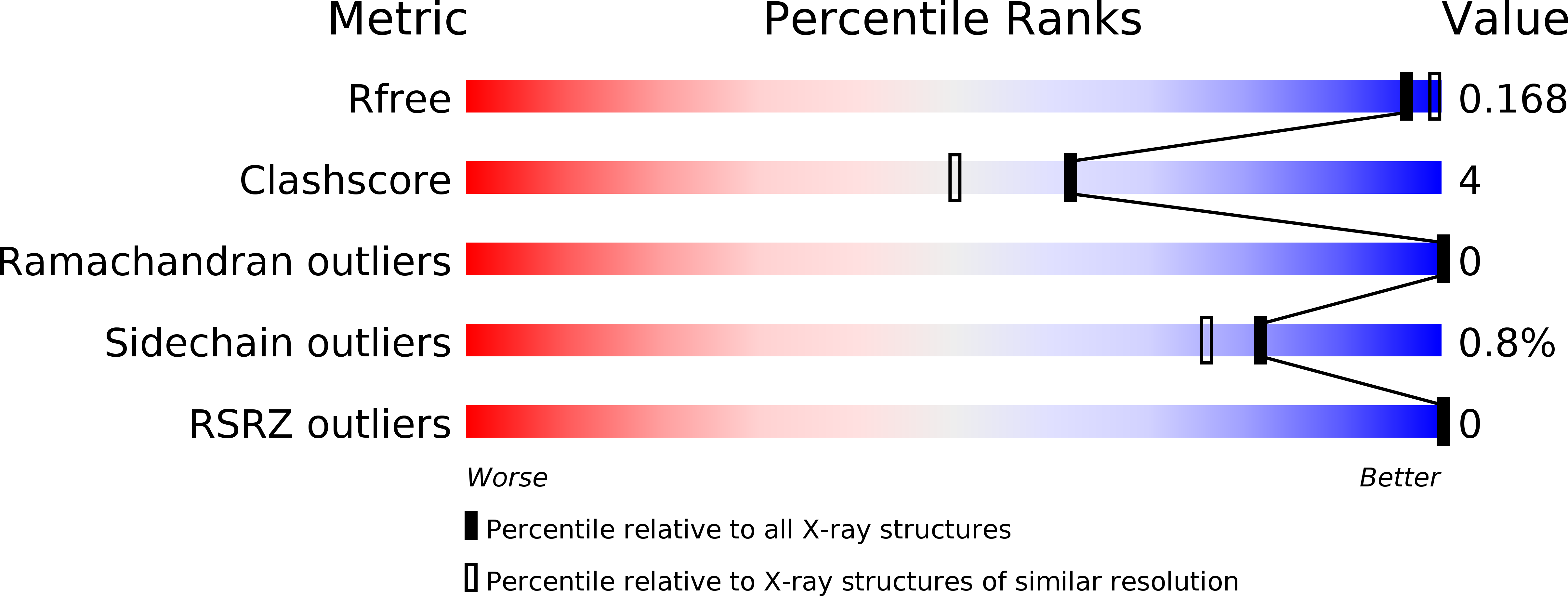
3WHO image
Deposition Date 2013-08-30
Release Date 2014-07-02
Last Version Date 2024-11-13
Entry Detail
PDB ID:
3WHO
Keywords:
Title:
X-ray-Crystallographic Structure of an RNase Po1 Exhibiting Anti-tumor Activity
Biological Source:
Source Organism:
Host Organism:
Method Details:
Experimental Method:
Resolution:
1.85 Å
R-Value Free:
0.24
R-Value Work:
0.20
R-Value Observed:
0.20
Space Group:
P 31
Macromolecular Entities
Structures with similar UniProt ID
Protein Blast
Polymer Type:polypeptide(L)
Molecule:Guanyl-specific ribonuclease Po1
Chain IDs:A, B, C
Chain Length:101
Number of Molecules:3
Biological Source:Pleurotus ostreatus
Primary Citation
X-ray crystallographic structure of RNase Po1 that exhibits anti-tumor activity.
Biol.Pharm.Bull. 37 968 978 (2014)
PMID: 24882409 DOI: 10.1248/bpb.b13-00929

Abstact

RNase Po1 is a guanylic acid-specific ribonuclease member of the RNase T1 family from Pleurotus ostreatus. We previously reported that RNase Po1 inhibits the proliferation of human tumor cells, yet RNase T1 and other T1 family RNases are non-toxic. We determined the three-dimensional X-ray structure of RNase Po1 and compared it with that of RNase T1. The catalytic sites are conserved. However, there are three disulfide bonds, one more than in RNase T1. One of the additional disulfide bond is in the catalytic and binding site of RNase Po1, and makes RNase Po1 more stable than RNase T1. A comparison of the electrostatic potential of the molecular surfaces of these two proteins shows that RNase T1 is anionic whereas RNase Po1 is cationic, so RNase Po1 might bind to the plasma membrane electrostatically. We suggest that the structural stability and cationic character of RNase Po1 are critical to the anti-cancer properties of the protein.

Legend

Protein

Chemical

Disease

Primary Citation of related structures