3WHC image
Entry Detail
PDB ID:
3WHC
Keywords:
Title:
Crystal structure of a transcriptional regulator FadR from Bacillus subtilis in complex with stearoyl-CoA
Biological Source:
Source Organism:
Host Organism:
PDB Version:
Deposition Date:
2013-08-23
Release Date:
2014-03-19
Method Details:
Experimental Method:
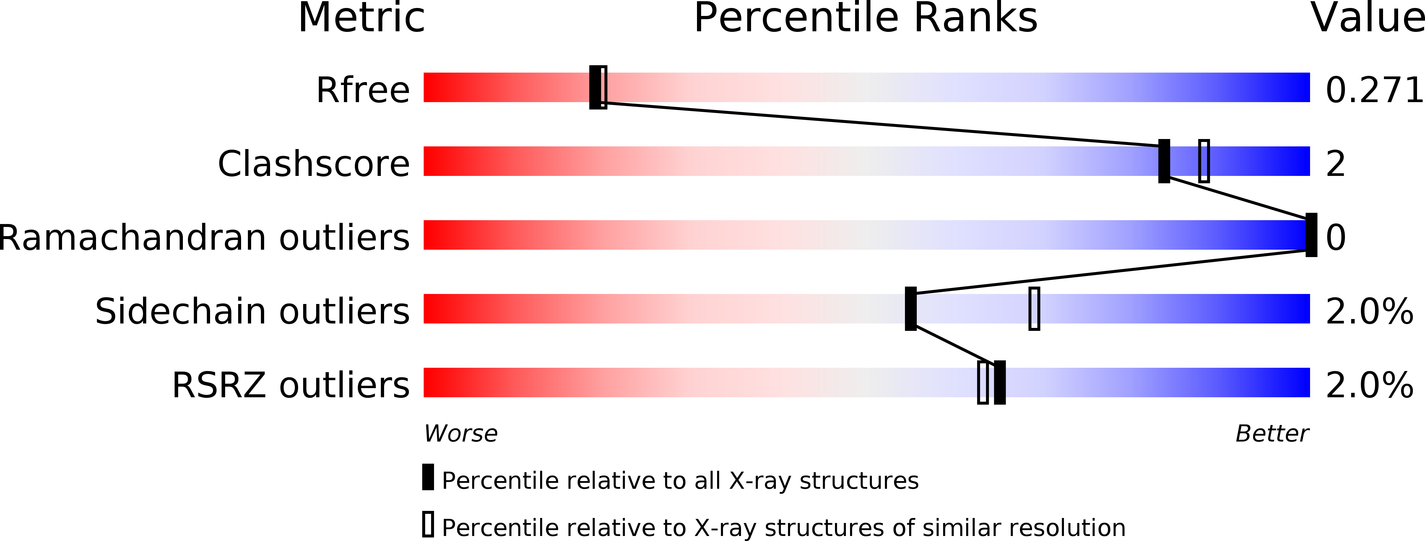
Resolution:
2.20 Å
R-Value Free:
0.27
R-Value Work:
0.23
R-Value Observed:
0.24
Space Group:
P 21 21 21
Macromolecular Entities
Polymer Type:polypeptide(L)
Description:Fatty acid metabolism regulator protein
Chain IDs:A, B, C, D, E, F
Chain Length:194
Number of Molecules:6
Biological Source:Bacillus subtilis
Ligand Molecules
Primary Citation
Structural characterization of a ligand-bound form of Bacillus subtilis FadR involved in the regulation of fatty acid degradation.
Proteins 82 1301 1310 (2014)
PMID: 24356978 DOI: 10.1002/prot.24496

Abstact

Bacillus subtilis FadR (FadR(Bs)), a member of the TetR family of bacterial transcriptional regulators, represses five fad operons including 15 genes, most of which are involved in β-oxidation of fatty acids. FadR(Bs) binds to the five FadR(Bs) boxes in the promoter regions and the binding is specifically inhibited by long-chain (C14-C20) acyl-CoAs, causing derepression of the fad operons. To elucidate the structural mechanism of this regulator, we have determined the crystal structures of FadR(Bs) proteins prepared with and without stearoyl(C18)-CoA. The crystal structure without adding any ligand molecules unexpectedly includes one small molecule, probably dodecyl(C12)-CoA derived from the Escherichia coli host, in its homodimeric structure. Also, we successfully obtained the structure of the ligand-bound form of the FadR(Bs) dimer by co-crystallization, in which two stearoyl-CoA molecules are accommodated, with the binding mode being essentially equivalent to that of dodecyl-CoA. Although the acyl-chain-binding cavity of FadR(Bs) is mainly hydrophobic, a hydrophilic patch encompasses the C1-C10 carbons of the acyl chain. This accounts for the previous report that the DNA binding of FadR(Bs) is specifically inhibited by the long-chain acyl-CoAs but not by the shorter ones. Structural comparison of the ligand-bound and unliganded subunits of FadR(Bs) revealed three regions around residues 21-31, 61-76, and 106-119 that were substantially changed in response to the ligand binding, and particularly with respect to the movements of Leu108 and Arg109. Site-directed mutagenesis of these residues revealed that Arg109, but not Leu108, is a key residue for maintenance of the DNA-binding affinity of FadR(Bs).

Legend

Protein

Chemical

Disease

Primary Citation of related structures