
Deposition Date
2013-08-21
Release Date
2014-09-03
Last Version Date
2023-11-08
Entry Detail
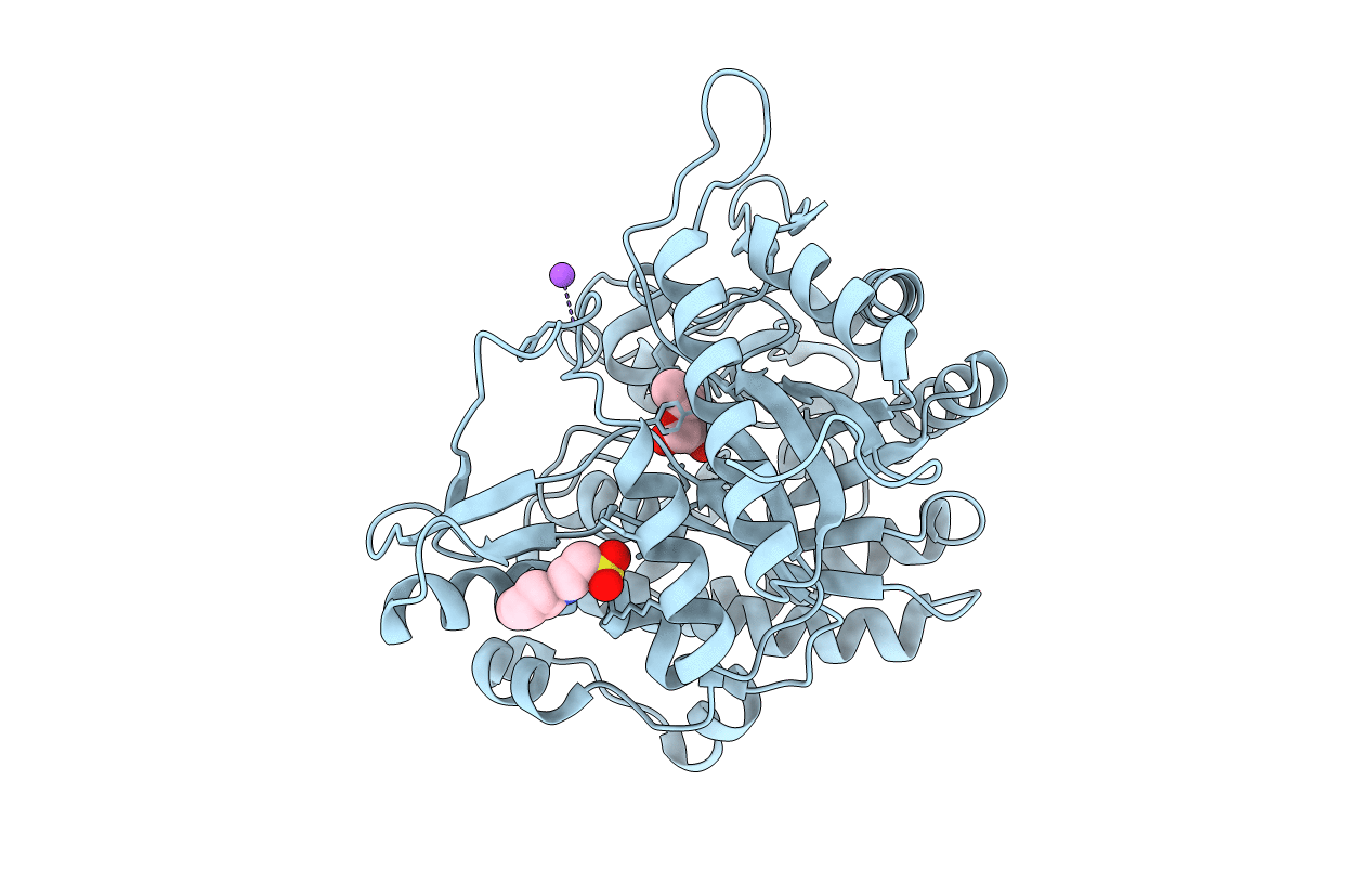
PDB ID:
3WH7
Keywords:
Title:
Crystal structure of GH1 beta-glucosidase Td2F2 L-fucose complex
Biological Source:
Source Organism(s):
metagenomes (Taxon ID: 408169)
Expression System(s):
Method Details:
Experimental Method:
Resolution:
1.10 Å
R-Value Free:
0.13
R-Value Work:
0.11
R-Value Observed:
0.11
Space Group:
P 21 21 21
