3WFU image
Deposition Date 2013-07-23
Release Date 2014-12-03
Last Version Date 2024-03-20
Entry Detail
PDB ID:
3WFU
Title:
Crystal structure of horse heart myoglobin reconstituted with cobalt(I) tetradehydrocorrin
Biological Source:
Source Organism:
Equus caballus (Taxon ID: 9796)
Method Details:
Experimental Method:
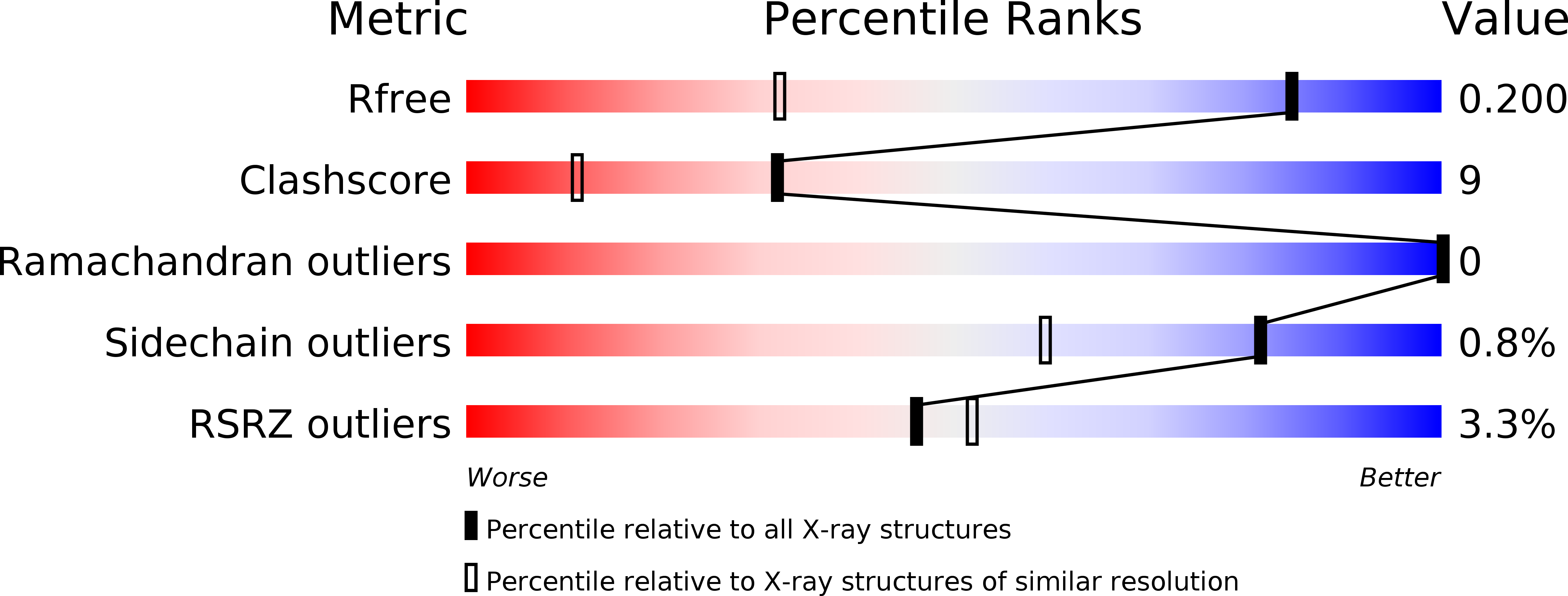
Resolution:
1.35 Å
R-Value Free:
0.19
R-Value Work:
0.14
R-Value Observed:
0.14
Space Group:
P 1 21 1
Macromolecular Entities
Polymer Type:polypeptide(L)
Molecule:Myoglobin
Gene (Uniprot):MB
Chain IDs:A
Chain Length:153
Number of Molecules:1
Biological Source:Equus caballus
Primary Citation
Co(II)/Co(I) reduction-induced axial histidine-flipping in myoglobin reconstituted with a cobalt tetradehydrocorrin as a methionine synthase model.
Chem.Commun.(Camb.) 50 12560 12563 (2014)
PMID: 25197974 DOI: 10.1039/c4cc05448b

Abstact

A conjugate between apomyoglobin and cobalt tetradehydrocorrin was prepared to replicate the coordination behavior of cob(I)alamin in methionine synthase. X-ray crystallography reveals that the tetra-coordinated Co(I) species is formed through the cleavage of the axial Co-His93 ligation after the reduction of the penta-coordinated Co(II) cofactor in the heme pocket.

Legend

Protein

Chemical

Disease

Primary Citation of related structures