Deposition Date
2013-07-03
Release Date
2014-01-01
Last Version Date
2023-11-08
Entry Detail
PDB ID:
3WEC
Keywords:
Title:
Structure of P450 RauA (CYP1050A1) complexed with a biosynthetic intermediate of aurachin RE
Biological Source:
Source Organism(s):
Rhodococcus erythropolis (Taxon ID: 1833)
Expression System(s):
Method Details:
Experimental Method:
Resolution:
2.19 Å
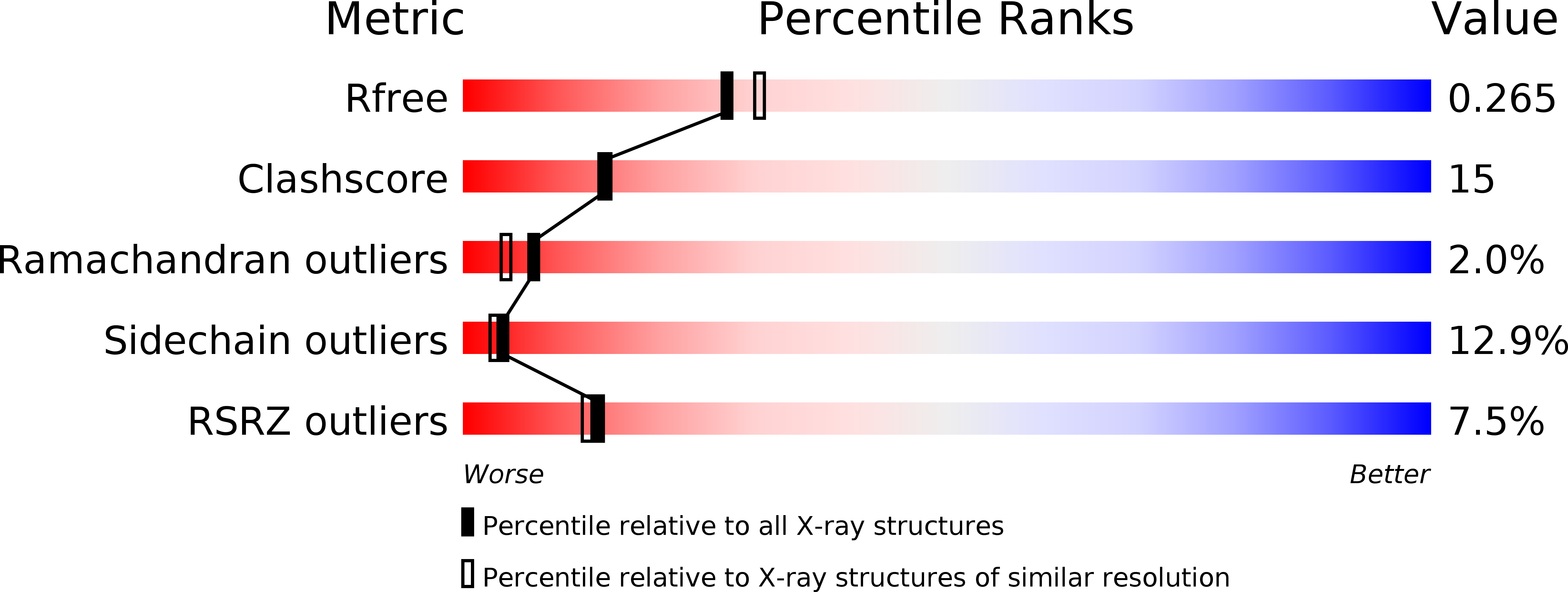
R-Value Free:
0.26
R-Value Work:
0.21
R-Value Observed:
0.21
Space Group:
P 1 21 1