3WDR image
Deposition Date 2013-06-20
Release Date 2014-03-05
Last Version Date 2024-04-03
Entry Detail
PDB ID:
3WDR
Keywords:
Title:
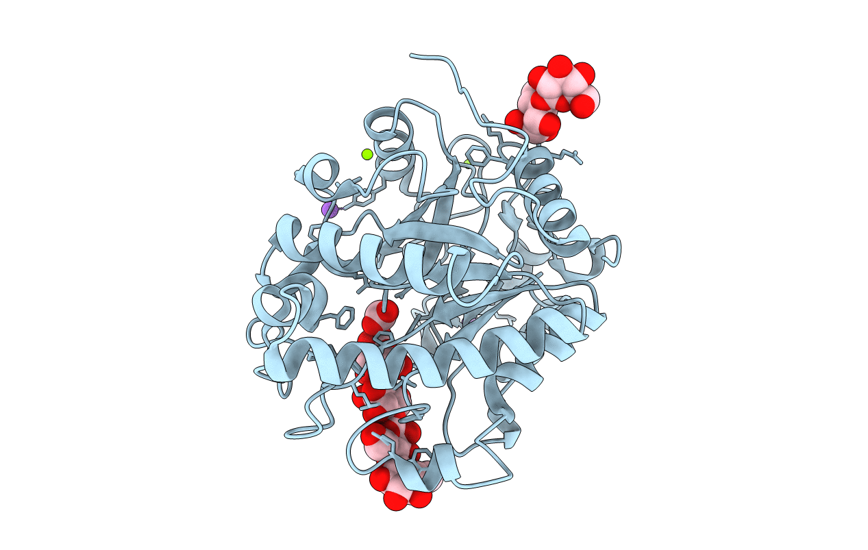
Crystal structure of beta-mannanase from a symbiotic protist of the termite Reticulitermes speratus complexed with gluco-manno-oligosaccharide
Biological Source:
Host Organism:
Method Details:
Experimental Method:
Resolution:
1.40 Å
R-Value Free:
0.15
R-Value Work:
0.13
R-Value Observed:
0.13
Space Group:
P 1 21 1
Macromolecular Entities
Polymer Type:polypeptide(L)
Molecule:Beta-mannanase
Gene (Uniprot):RsManC
Chain IDs:A
Chain Length:355
Number of Molecules:1
Biological Source:Symbiotic protist of Reticulitermes speratus
Modified Residue
Compound ID Chain ID Parent Comp ID Details 2D Image
SME A MET METHIONINE SULFOXIDE
Primary Citation
Structural and Biochemical Analyses of Glycoside Hydrolase Family 26 beta-Mannanase from a Symbiotic Protist of the Termite Reticulitermes speratus
J.Biol.Chem. 289 10843 10852 (2014)
PMID: 24570006 DOI: 10.1074/jbc.M114.555383

Abstact

Termites and their symbiotic protists have established a prominent dual lignocellulolytic system, which can be applied to the biorefinery process. One of the major components of lignocellulose from conifers is glucomannan, which comprises a heterogeneous combination of β-1,4-linked mannose and glucose. Mannanases are known to hydrolyze the internal linkage of the glucomannan backbone, but the specific mechanism by which they recognize and accommodate heteropolysaccharides is currently unclear. Here, we report biochemical and structural analyses of glycoside hydrolase family 26 mannanase C (RsMan26C) from a symbiotic protist of the termite Reticulitermes speratus. RsMan26C was characterized based on its catalytic efficiency toward glucomannan, compared with pure mannan. The crystal structure of RsMan26C complexed with gluco-manno-oligosaccharide(s) explained its specificities for glucose and mannose at subsites -5 and -2, respectively, in addition to accommodation of both glucose and mannose at subsites -3 and -4. RsMan26C has a long open cleft with a hydrophobic platform of Trp(94) at subsite -5, facilitating enzyme binding to polysaccharides. Notably, a unique oxidized Met(85) specifically interacts with the equatorial O-2 of glucose at subsite -3. Our results collectively indicate that specific recognition and accommodation of glucose at the distal negative subsites confers efficient degradation of the heteropolysaccharide by mannanase.

Legend

Protein

Chemical

Disease

Primary Citation of related structures