Deposition Date
2013-06-19
Release Date
2013-10-30
Last Version Date
2023-11-08
Entry Detail
PDB ID:
3WDN
Keywords:
Title:
High-resolution X-ray crystal structure of bovine H-protein using a high-pressure cryocooling method
Biological Source:
Source Organism(s):
Bos taurus (Taxon ID: 9913)
Expression System(s):
Method Details:
Experimental Method:
Resolution:
0.86 Å
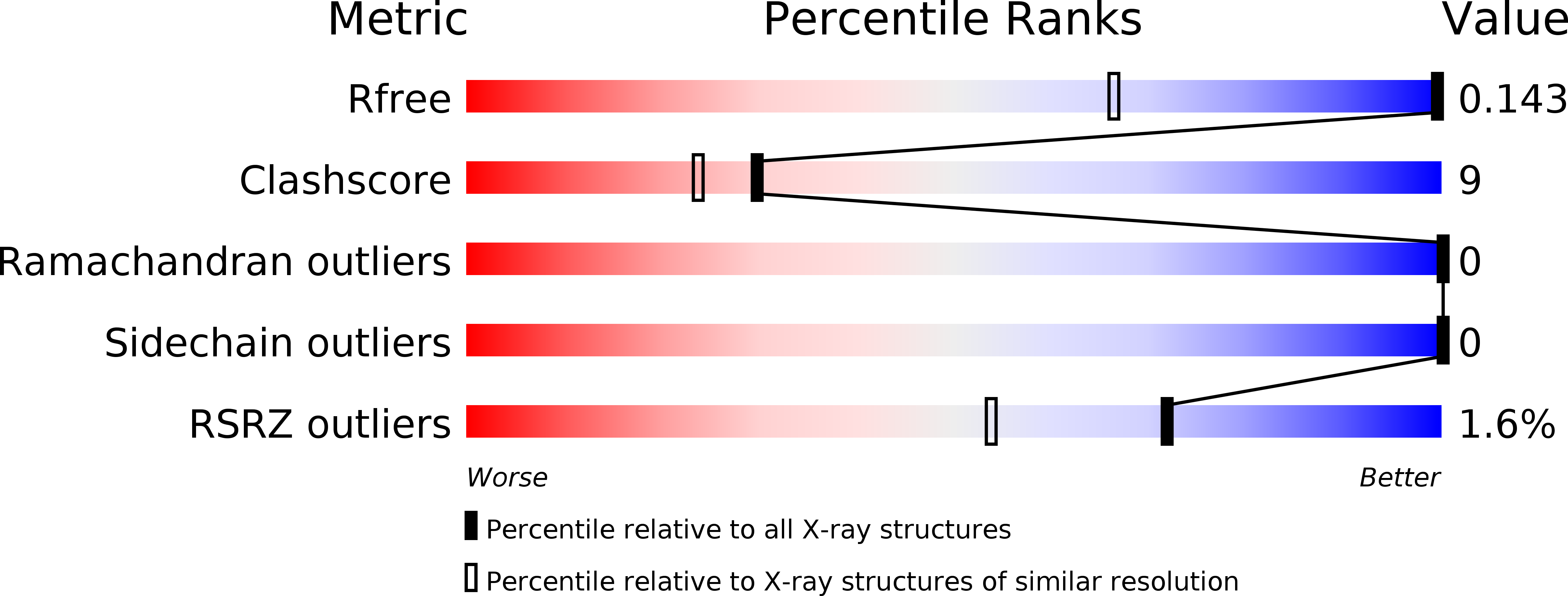
R-Value Free:
0.13
R-Value Work:
0.11
Space Group:
C 1 2 1