3WC4 image
Deposition Date 2013-05-24
Release Date 2013-10-30
Last Version Date 2023-11-08
Entry Detail
PDB ID:
3WC4
Keywords:
Title:
Crystal structure of UDP-glucose: anthocyanidin 3-O-glucosyltransferase from Clitoria ternatea
Biological Source:
Source Organism:
Host Organism:
Method Details:
Experimental Method:
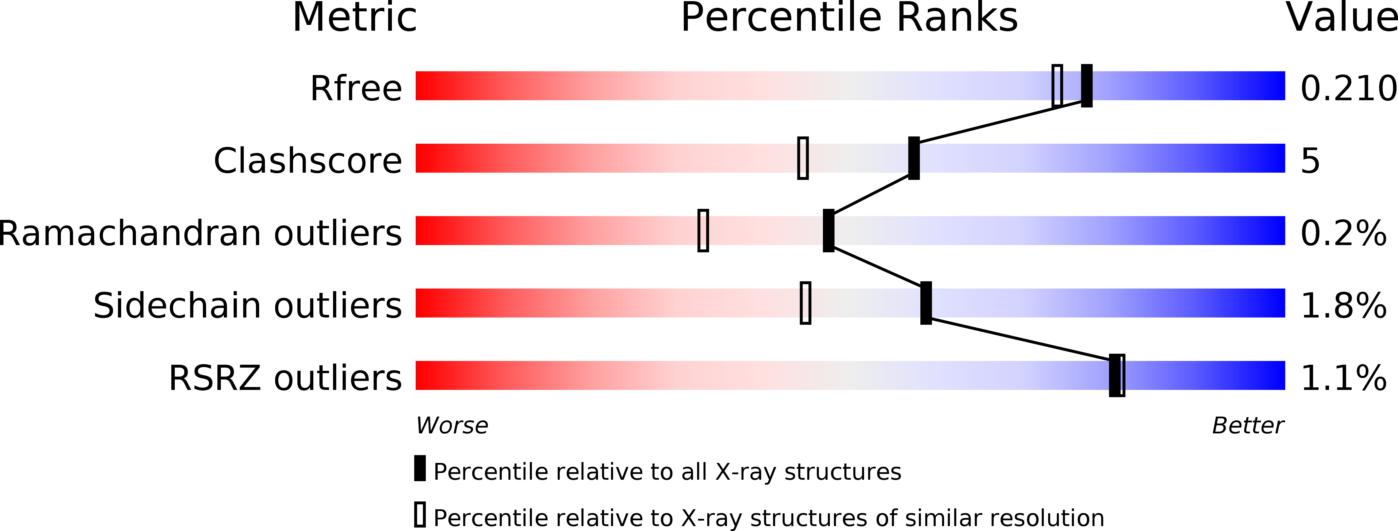
Resolution:
1.85 Å
R-Value Free:
0.21
R-Value Work:
0.17
R-Value Observed:
0.17
Space Group:
P 1 21 1
Macromolecular Entities
Structures with similar UniProt ID
Protein Blast
Polymer Type:polypeptide(L)
Molecule:UDP-glucose:anthocyanidin 3-O-glucosyltransferase
Gene (Uniprot):Ct3GT-A
Chain IDs:A
Chain Length:446
Number of Molecules:1
Biological Source:Clitoria ternatea
Primary Citation
Crystal structure of UDP-glucose:anthocyanidin 3-O-glucosyltransferase from Clitoria ternatea
J.SYNCHROTRON RADIAT. 20 894 898 (2013)
PMID: 24121335 DOI: 10.1107/S0909049513020712

Abstact

Flowers of the butterfly pea (Clitoria ternatea) accumulate a group of polyacylated anthocyanins, named ternatins, in their petals. The first step in ternatin biosynthesis is the transfer of glucose from UDP-glucose to anthocyanidins such as delphinidin, a reaction catalyzed in C. ternatea by UDP-glucose:anthocyanidin 3-O-glucosyltransferase (Ct3GT-A; AB185904). To elucidate the structure-function relationship of Ct3GT-A, recombinant Ct3GT-A was expressed in Escherichia coli and its tertiary structure was determined to 1.85 Å resolution by using X-ray crystallography. The structure of Ct3GT-A shows a common folding topology, the GT-B fold, comprised of two Rossmann-like β/α/β domains and a cleft located between the N- and C-domains containing two cavities that are used as binding sites for the donor (UDP-Glc) and acceptor substrates. By comparing the structure of Ct3GT-A with that of the flavonoid glycosyltransferase VvGT1 from red grape (Vitis vinifera) in complex with UDP-2-deoxy-2-fluoro glucose and kaempferol, locations of the catalytic His-Asp dyad and the residues involved in recognizing UDP-2-deoxy-2-fluoro glucose were essentially identical in Ct3GT-A, but certain residues of VvGT1 involved in binding kaempferol were found to be substituted in Ct3GT-A. These findings are important for understanding the differentiation of acceptor-substrate recognition in these two enzymes.

Legend

Protein

Chemical

Disease

Primary Citation of related structures