3WBD image
Deposition Date 2013-05-14
Release Date 2013-10-16
Last Version Date 2024-11-20
Entry Detail
PDB ID:
3WBD
Keywords:
Title:
Crystal structure of anti-polysialic acid antibody single chain Fv fragment (mAb735) complexed with octasialic acid
Biological Source:
Source Organism:
Mus musculus (Taxon ID: 10090)
Host Organism:
Method Details:
Experimental Method:
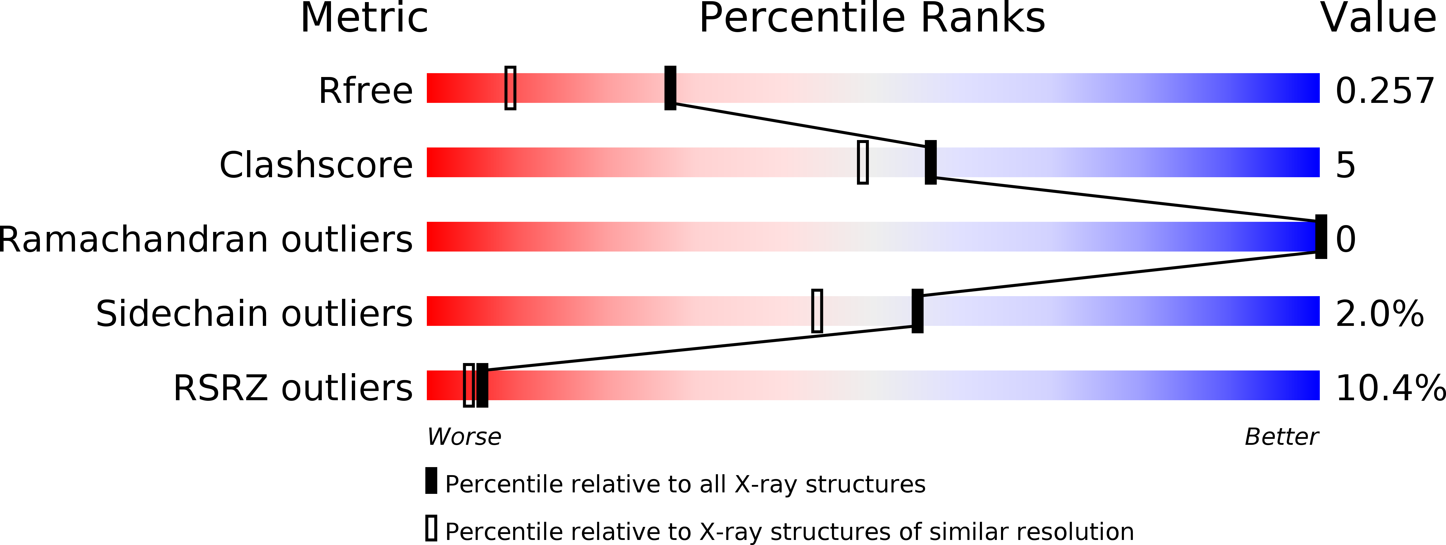
Resolution:
1.80 Å
R-Value Free:
0.25
R-Value Work:
0.23
R-Value Observed:
0.23
Space Group:
P 64
Macromolecular Entities
Polymer Type:polypeptide(L)
Molecule:single chain Fv fragment of mAb735
Chain IDs:A, B
Chain Length:246
Number of Molecules:2
Biological Source:Mus musculus
Ligand Molecules
Primary Citation
Crystal structure of anti-polysialic acid antibody single chain Fv fragment complexed with octasialic acid: insight into the binding preference for polysialic acid.
J.Biol.Chem. 288 33784 33796 (2013)
PMID: 24100042 DOI: 10.1074/jbc.M113.496224

Abstact

Polysialic acid is a linear homopolymer of α2-8-linked sialic acids attached mainly onto glycoproteins. Cell surface polysialic acid plays roles in cell adhesion and differentiation events in a manner that is often dependent on the degree of polymerization (DP). Anti-oligo/polysialic acid antibodies have DP-dependent antigenic specificity, and such antibodies are widely utilized in biological studies for detecting and distinguishing between different oligo/polysialic acids. A murine monoclonal antibody mAb735 has a unique preference for longer polymers of polysialic acid (DP >10), yet the mechanism of recognition at the atomic level remains unclear. Here, we report the crystal structure of mAb735 single chain variable fragment (scFv735) in complex with octasialic acid at 1.8 Å resolution. In the asymmetric unit, two scFv735 molecules associate with one octasialic acid. In both complexes of the unit, all the complementarity-determining regions except for L3 interact with three consecutive sialic acid residues out of the eight. A striking feature of the complex is that 11 ordered water molecules bridge the gap between antibody and ligand, whereas the direct antibody-ligand interaction is less extensive. The dihedral angles of the trisialic acid unit directly interacting with scFv735 are not uniform, indicating that mAb735 does not strictly favor the previously proposed helical conformation. Importantly, both reducing and nonreducing ends of the bound ligand are completely exposed to solvent. We suggest that mAb735 gains its apparent high affinity for a longer polysialic acid chain by recognizing every three sialic acid units in a paired manner.

Legend

Protein

Chemical

Disease

Primary Citation of related structures