Deposition Date
2013-05-10
Release Date
2014-01-29
Last Version Date
2023-11-08
Entry Detail
PDB ID:
3WAZ
Keywords:
Title:
Crystal structure of a restriction enzyme PabI in complex with DNA
Biological Source:
Source Organism(s):
Pyrococcus abyssi (Taxon ID: 272844)
synthetic construct (Taxon ID: 32630)
synthetic construct (Taxon ID: 32630)
Expression System(s):
Method Details:
Experimental Method:
Resolution:
3.00 Å
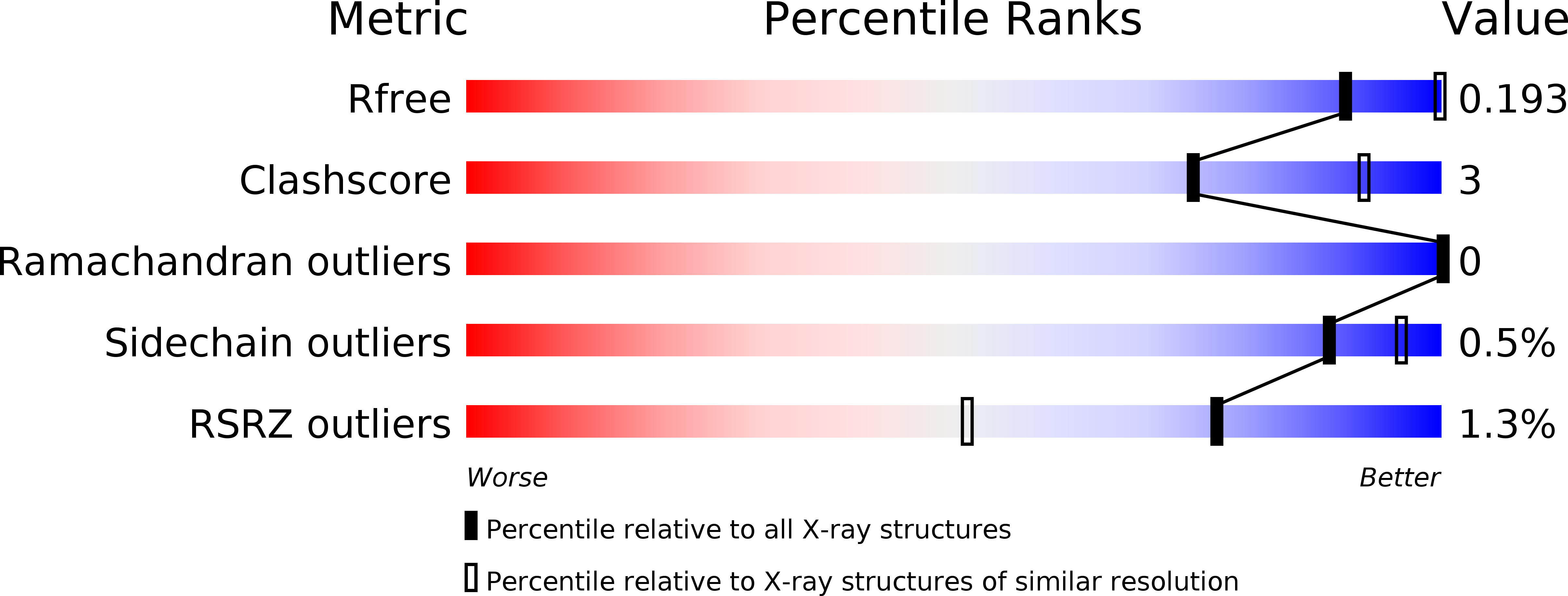
R-Value Free:
0.19
R-Value Work:
0.16
R-Value Observed:
0.17
Space Group:
I 2 2 2