Deposition Date
2013-05-03
Release Date
2013-10-30
Last Version Date
2023-11-08
Entry Detail
PDB ID:
3WAJ
Keywords:
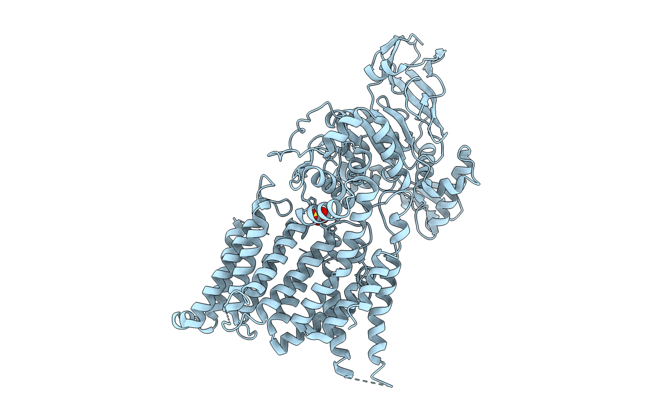
Title:
Crystal structure of the Archaeoglobus fulgidus oligosaccharyltransferase (O29867_ARCFU) complex with Zn and sulfate
Biological Source:
Source Organism(s):
Archaeoglobus fulgidus (Taxon ID: 224325)
Expression System(s):
Method Details:
Experimental Method:
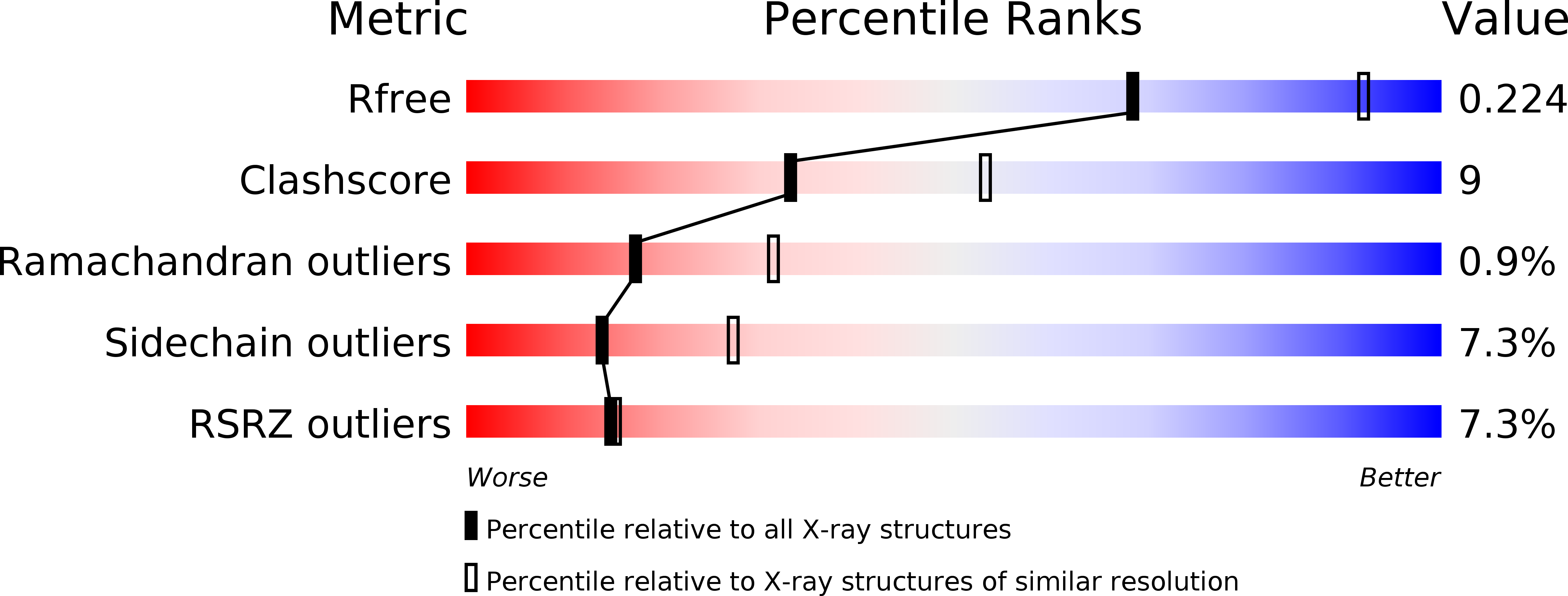
Resolution:
2.50 Å
R-Value Free:
0.21
R-Value Work:
0.18
R-Value Observed:
0.18
Space Group:
C 1 2 1