
Deposition Date
2013-05-03
Release Date
2013-07-17
Last Version Date
2023-11-08
Entry Detail
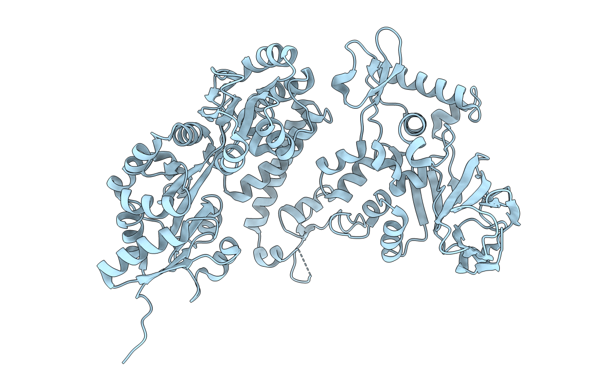
PDB ID:
3WAI
Keywords:
Title:
Crystal structure of the C-terminal globular domain of oligosaccharyltransferase (AfAglB-L, O29867_ARCFU) from Archaeoglobus fulgidus as a MBP fusion
Biological Source:
Source Organism(s):
Escherichia coli (Taxon ID: 83333)
Archaeoglobus fulgidus (Taxon ID: 224325)
Archaeoglobus fulgidus (Taxon ID: 224325)
Expression System(s):
Method Details:
Experimental Method:
Resolution:
1.90 Å
R-Value Free:
0.20
R-Value Work:
0.16
R-Value Observed:
0.16
Space Group:
C 1 2 1
