
Deposition Date
2013-04-22
Release Date
2014-07-02
Last Version Date
2024-10-30
Entry Detail
PDB ID:
3WA1
Keywords:
Title:
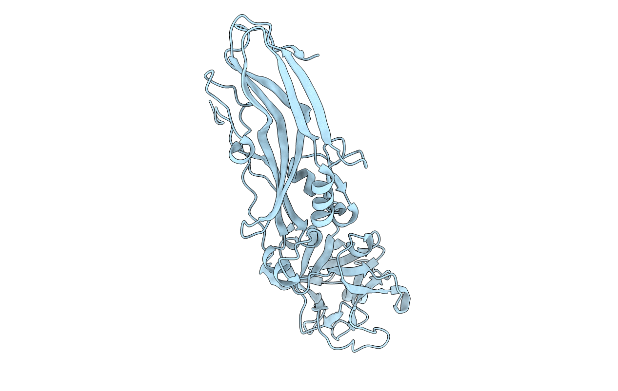
Crystal structure of BinB: A receptor binding component of the binary toxin from Lysinibacillus sphaericus
Biological Source:
Source Organism(s):
Lysinibacillus sphaericus (Taxon ID: 1421)
Expression System(s):
Method Details:
Experimental Method:
Resolution:
1.75 Å
R-Value Free:
0.21
R-Value Work:
0.17
R-Value Observed:
0.17
Space Group:
P 62 2 2
