3W9Y image
Deposition Date 2013-04-18
Release Date 2013-06-26
Last Version Date 2024-11-06
Entry Detail
PDB ID:
3W9Y
Title:
Crystal structure of the human DLG1 guanylate kinase domain
Biological Source:
Source Organism:
Homo sapiens (Taxon ID: 9606)
Host Organism:
Method Details:
Experimental Method:
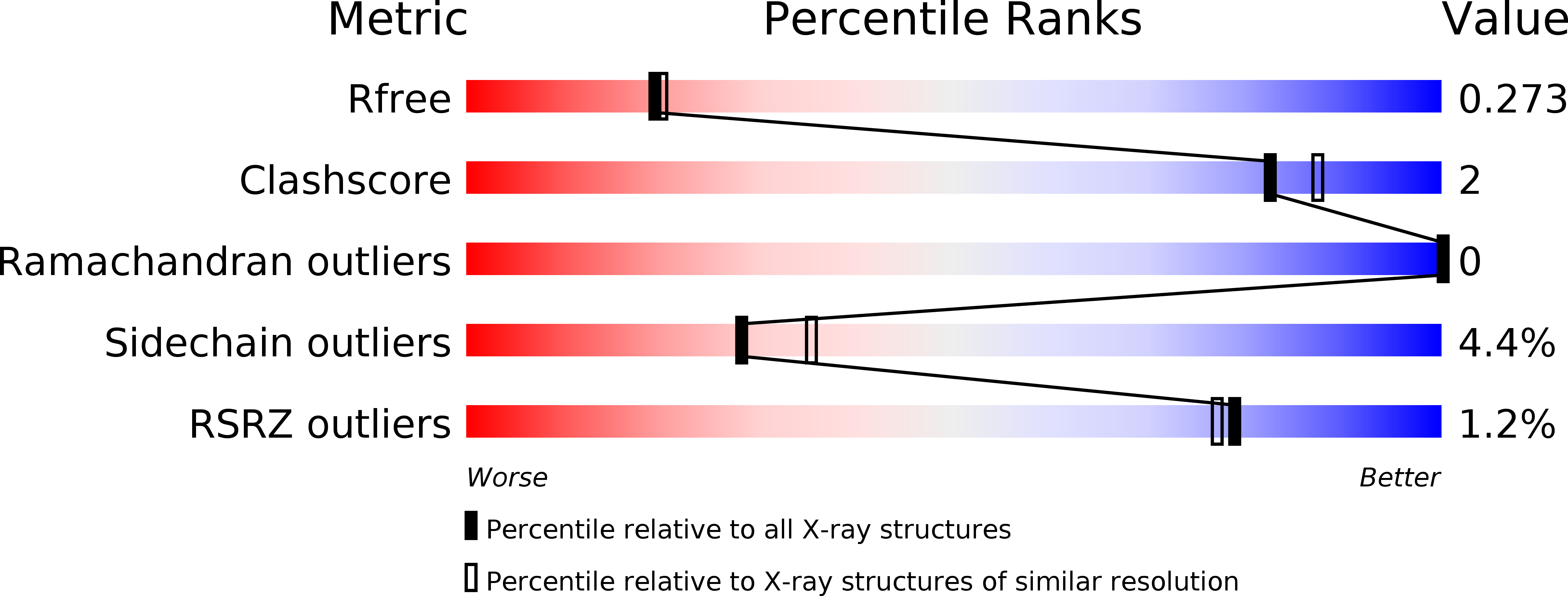
Resolution:
2.20 Å
R-Value Free:
0.26
R-Value Work:
0.20
R-Value Observed:
0.21
Space Group:
P 65 2 2
Macromolecular Entities
Structures with similar UniProt ID
Protein Blast
Polymer Type:polypeptide(L)
Molecule:Disks large homolog 1
Chain IDs:A
Chain Length:200
Number of Molecules:1
Biological Source:Homo sapiens
Modified Residue
Compound ID Chain ID Parent Comp ID Details 2D Image
MSE A MET SELENOMETHIONINE
Primary Citation
Crystal structure of the guanylate kinase domain from discs large homolog 1 (DLG1/SAP97)
Biochem.Biophys.Res.Commun. 435 334 338 (2013)
PMID: 23624197 DOI: 10.1016/j.bbrc.2013.04.056

Abstact

Discs large homolog 1 (DLG1/SAP97) is involved in the development and regulation of neuronal and immunological synapses. DLG1 is a member of the membrane associated guanylate kinase (MAGUK) family of proteins, which function as molecular scaffolds. The C-terminal guanylate kinase (GK) domain of DLG1 binds peptides with a phosphorylated serine residue. In this study, we solved the crystal structure of the GK domain of human DLG1. The C-terminal tail of DLG1 is bound to the peptide-binding site of an adjacent symmetry-related DLG1 GK molecule. The binding direction of the C-terminal tail to the peptide-binding site is opposite to that of the phosphorylated LGN peptide in complex with the rat DLG1 GK domain. The C-terminal tail forms a 310 helix, which is also different from the conformation of the phosphorylated LGN peptide. Nevertheless, the side chain interactions of the C-terminal tail with the DLG1 GK domain are similar to those of the phosphorylated LGN peptide.

Legend

Protein

Chemical

Disease

Primary Citation of related structures