3W82 image
Deposition Date 2013-03-11
Release Date 2013-08-21
Last Version Date 2024-10-30
Entry Detail
PDB ID:
3W82
Keywords:
Title:
Human alpha-L-iduronidase in complex with iduronic acid
Biological Source:
Source Organism:
Homo sapiens (Taxon ID: 9606)
Host Organism:
Method Details:
Experimental Method:
Resolution:
2.76 Å
R-Value Free:
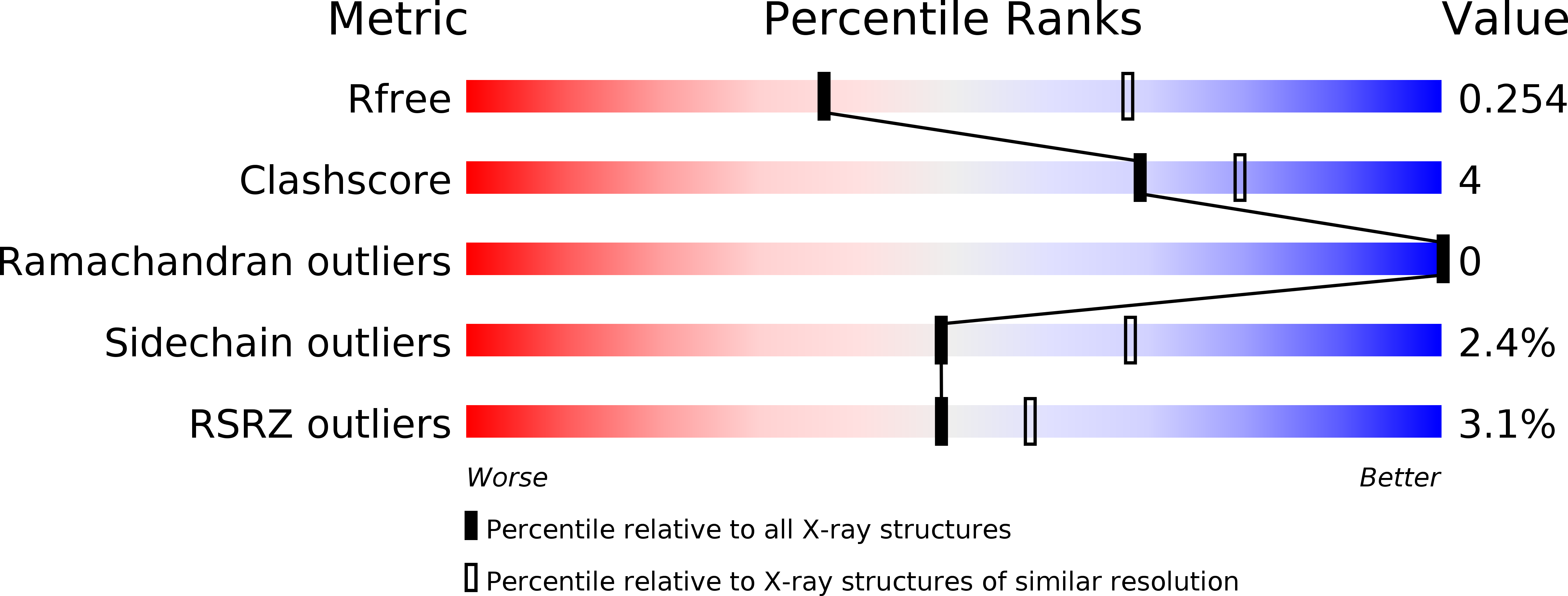
0.25
R-Value Work:
0.19
R-Value Observed:
0.19
Space Group:
H 3
Macromolecular Entities
Structures with similar UniProt ID
Protein Blast
Polymer Type:polypeptide(L)
Molecule:Alpha-L-iduronidase
Gene (Uniprot):IDUA
Chain IDs:A, B
Chain Length:627
Number of Molecules:2
Biological Source:Homo sapiens
Modified Residue
Compound ID Chain ID Parent Comp ID Details 2D Image
ASN B ASN GLYCOSYLATION SITE
Primary Citation
Human alpha-L-iduronidase uses its own N-glycan as a substrate-binding and catalytic module
Proc.Natl.Acad.Sci.USA 110 14628 14633 (2013)
PMID: 23959878 DOI: 10.1073/pnas.1306939110

Abstact

N-glycosylation is a major posttranslational modification that endows proteins with various functions. It is established that N-glycans are essential for the correct folding and stability of some enzymes; however, the actual effects of N-glycans on their activities are poorly understood. Here, we show that human α-l-iduronidase (hIDUA), of which a dysfunction causes accumulation of dermatan/heparan sulfate leading to mucopolysaccharidosis type I, uses its own N-glycan as a substrate binding and catalytic module. Structural analysis revealed that the mannose residue of the N-glycan attached to N372 constituted a part of the substrate-binding pocket and interacted directly with a substrate. A deglycosylation study showed that enzyme activity was highly correlated with the N-glycan attached to N372. The kinetics of native and deglycosylated hIDUA suggested that the N-glycan is also involved in catalytic processes. Our study demonstrates a previously unrecognized function of N-glycans.

Legend

Protein

Chemical

Disease

Primary Citation of related structures